|
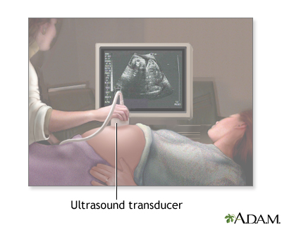
As you lie on an exam table, a sonographer coats your belly with a slick -- and possibly cold -- gel. Next, he moves a transducer, a hand-held device shaped like a microphone, over your belly. You can see the resulting images on a nearby computer screen.
|
|
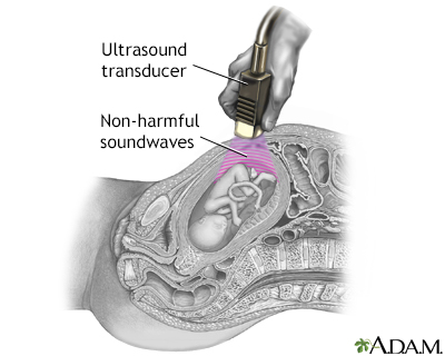
The transducer emits inaudible sound waves, which fan out as they travel through your abdomen. When they hit dense structures like the fetus and the wall of your uterus, the sound waves bounce back to the transducer and are translated into a visual image by the computer.
|
|
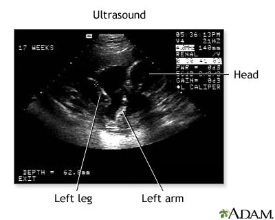
Don't get your hopes up too much about this first, fleeting look at your baby. The black-and-white image you see on the computer screen is grainy, shadowy, and may look more like a test pattern than a baby-to-be. Your sonographer will walk you through what you're seeing by pointing out the fetus' developing heart, limbs, and head.
|
|
Review Date: 12/9/2012 Reviewed By: Irina Burd, MD, PhD, Maternal Fetal Medicine, Johns Hopkins University, Baltimore, MD. Review provided by VeriMed Healthcare Network.
The information provided herein should not be used during any medical emergency or for the diagnosis or treatment of any medical condition. A licensed medical professional should be consulted for diagnosis and treatment of any and all medical conditions. Links to other sites are provided for information only -- they do not constitute endorsements of those other sites. No warranty of any kind, either expressed or implied, is made as to the accuracy, reliability, timeliness, or correctness of any translations made by a third-party service of the information provided herein into any other language. © 1997-
A.D.A.M., a business unit of Ebix, Inc. Any duplication or distribution of the information contained herein is strictly prohibited.
|