Pneumothorax - series
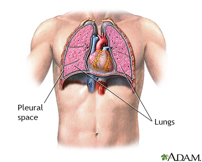
Normal
The lungs are paired organs that lie in the thoracic cavity. The lungs extract oxygen from inhaled air and transport the oxygen to the blood. Surrounding the lungs is a very thin space called the pleural space. The pleural space is usually extremely thin, and filled with a small amount of fluid.
Indications
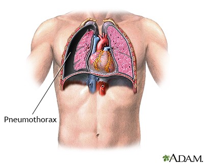
If air enters the pleural space, the lung will collapse. This is called a pneumothorax. If the chest wall is penetrated, which may occur as a result of an injury, air can enter the pleural space from the outside. Air can also enter from the inside, from the lung itself, if the lung is torn or ruptured. One of the most common causes of spontaneous non-traumatic pneumothorax is a pulmonary bleb. This is a weakness and out-pouching of the lung tissue, which can rupture. This introduces air into the pleural space.
Incision
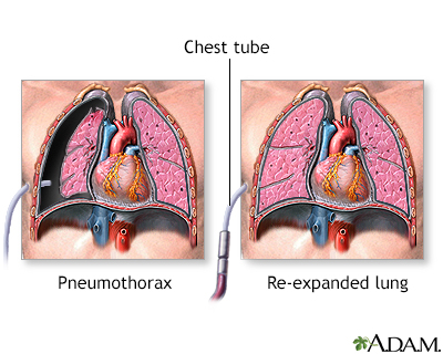
Pneumothorax can be life-threatening. The immediate treatment for pneumothorax is tube thoracostomy, or the insertion of a chest tube. A long, flexible, hollow, narrow tube is inserted through the ribs into the pleural splace, and the tube is attached to a suction device. This allows the air to be evacuated from the pleural space, and allows the lung to re-expand. Chest tubes are generally inserted using local anesthesia. The chest tube is left in place until the lung leak seals on its own; this usually occurs within two to five days.
Indications
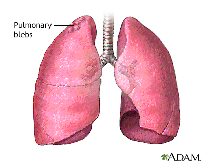
Pulmonary blebs are a common cause of spontaneous pneumothorax in young children and adults. Blebs are weakened out-pouchings in the upper lung, which can rupture, causing pneumothorax.
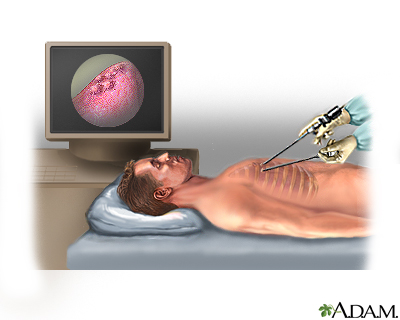
Procedure
Pulmonary blebs can be resected, preventing future pneumothorax. This is frequently done using a thoracoscopic surgical procedure. The patient is put to sleep using general anesthesia. Long, narrow instruments, including one with a camera on the end to allow for visualization of the interior of the thorax, are introduced through small incisions in the chest wall.
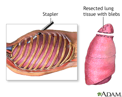
Aftercare
A stapling device is inserted into the chest during thoracoscopic surgery, and the segment of lung with blebs is stapled across and then removed. Most patients respond quite well to this procedure, and usually require one to three days in the hospital after surgery to recover. A chest tube is frequently left in place for one to two days after surgery to evacuate any residual air in the pleural space.
BACK TO TOP
Review Date: 1/2/2023
Reviewed By: Jesse Borke, MD, CPE, FAAEM, FACEP, Attending Physician at Kaiser Permanente, Orange County, CA. Also reviewed by David C. Dugdale, MD, Medical Director, Brenda Conaway, Editorial Director, and the A.D.A.M. Editorial team.

Health Content Provider
06/01/2025
|
A.D.A.M., Inc. is accredited by URAC, for Health Content Provider (www.urac.org). URAC's accreditation program is an independent audit to verify that A.D.A.M. follows rigorous standards of quality and accountability. A.D.A.M. is among the first to achieve this important distinction for online health information and services. Learn more about A.D.A.M.'s editorial policy, editorial process and privacy policy. A.D.A.M. is also a founding member of Hi-Ethics. This site complied with the HONcode standard for trustworthy health information from 1995 to 2022, after which HON (Health On the Net, a not-for-profit organization that promoted transparent and reliable health information online) was discontinued. |
The information provided herein should not be used during any medical emergency or for the diagnosis or treatment of any medical condition. A licensed medical professional should be consulted for diagnosis and treatment of any and all medical conditions. Links to other sites are provided for information only -- they do not constitute endorsements of those other sites. © 1997- 2025 A.D.A.M., a business unit of Ebix, Inc. Any duplication or distribution of the information contained herein is strictly prohibited.
 All rights reserved.