Retinal detachment repair - series
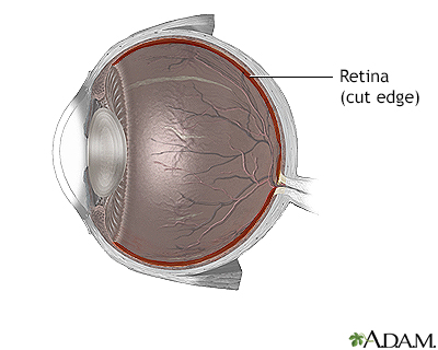
Normal anatomy
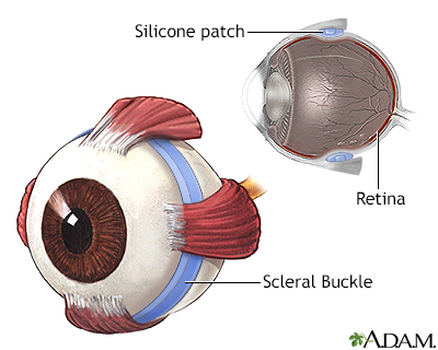
The retina is the internal layer of the eye that receives and transmits images that have passed through and been focused by the lens and cornea.
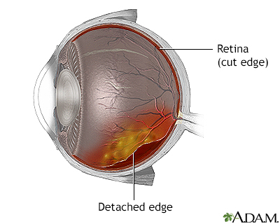
Indications
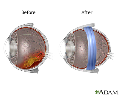
Retinal detachments are associated with a tear or hole in the retina through which the internal fluids of the eye may leak, causing separation of the retina from the underlying tissues. This is most often caused by trauma, and the risk of retinal detachment after minor trauma, such as a blow to the head, is increased in the elderly, and in patients with tumors or inflammation near the retina. In some cases, retinal detachment occurs in the absence of trauma. Symptoms of retinal detachment include bright flashes, floaters, or loss of part of the visual field. Emergency retinal detachment surgery is necessary to prevent vision loss.
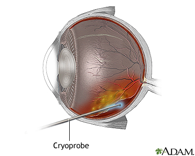
Procedure, part 1
The most common technique used to repair retinal detachment is called a scleral buckle.
Prior to performing a scleral buckle procedure, breaks and tears in the retina are closed. There are two major methods used to close breaks and tears in the retina. Cryopexy, uses an intensely cold probe (cryoprobe). This produces an inflammation that leads to formation of a scar which holds the retina to the underlying tissue.
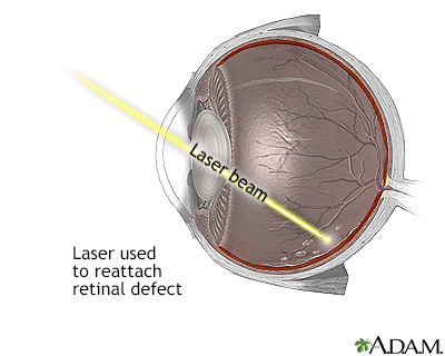
Procedure, part 2
A laser treatment (photocoagulation) can also be used to seals holes in the retina. The choice of cryopexy or photocoagulation is usually determined by the preference of the surgeon-both procedures are equally effective in most cases.
Procedure, part 3
After sealing breaks and tears in the retina with either laser or cryoporbe treatment, the scleral buckle is applied. This consists of a silicone patch wrapped around the eye, compressing the globe and elongating it slightly, thus pushing the retina up against the posterior aspect of the eye, and sealing the detachment. The silicone patch is usually left in place permanently, unless it causes problems later, such as infection.
Aftercare
Scleral buckling for detachment may require a few days in the hospital. Keep the head elevated at all times. Patients should not bend over or strain with lifting or bowel movements. Vigorous exercise should be avoided for 3 to 4 weeks.
BACK TO TOP
Review Date: 8/5/2024
Reviewed By: Franklin W. Lusby, MD, Ophthalmologist, Lusby Vision Institute, La Jolla, CA. Also reviewed by David C. Dugdale, MD, Medical Director, Brenda Conaway, Editorial Director, and the A.D.A.M. Editorial team.

Health Content Provider
06/01/2025
|
A.D.A.M., Inc. is accredited by URAC, for Health Content Provider (www.urac.org). URAC's accreditation program is an independent audit to verify that A.D.A.M. follows rigorous standards of quality and accountability. A.D.A.M. is among the first to achieve this important distinction for online health information and services. Learn more about A.D.A.M.'s editorial policy, editorial process and privacy policy. A.D.A.M. is also a founding member of Hi-Ethics. This site complied with the HONcode standard for trustworthy health information from 1995 to 2022, after which HON (Health On the Net, a not-for-profit organization that promoted transparent and reliable health information online) was discontinued. |
The information provided herein should not be used during any medical emergency or for the diagnosis or treatment of any medical condition. A licensed medical professional should be consulted for diagnosis and treatment of any and all medical conditions. Links to other sites are provided for information only -- they do not constitute endorsements of those other sites. © 1997- 2025 A.D.A.M., a business unit of Ebix, Inc. Any duplication or distribution of the information contained herein is strictly prohibited.
 All rights reserved.