Spinal fusion - series
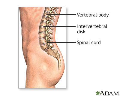
Normal anatomy
The vertebrae are the bones that make up the spinal column, which surrounds and protects the spinal cord. The intervertebral disks are soft tissues that sit between each vertebrae and act as cushions between vertebrae, and absorb energy while the spinal column flexes, extends, and twists. Nerves from the spinal cord exit the spinal column between each vertebra.
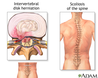
Indications
Spinal fusion is a surgical technique in which one or more vertebrae are fused together to stop the motion between them.
Spinal fusion may be recommended for:
- Abnormal curvature of the spine (scoliosis or kyphosis)
- Injury to the spinal vertebrae
- Protrusion of the cushioning disk between vertebrae (slipped disk, herniated nucleus pulposus)
- Weak or unstable spine caused by infections or tumors
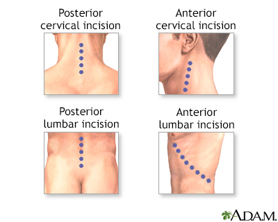
Incision
Different incisions are made depending on the area to be treated. The approach can be made either from the front (anterior), from the back (posterior), or both.
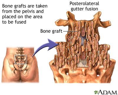
Procedure: Posterolateral gutter fusion
In a posterolateral gutter fusion procedure, the spine is approached from the back. Bone graft is taken from the pelvis and laid out in the posterolateral portion of the spine that is to be fused. The back muscles hold the graft in place until it fuses with the vertebrae. A fusion will setup within three months and will continue to get stronger for one to two years.
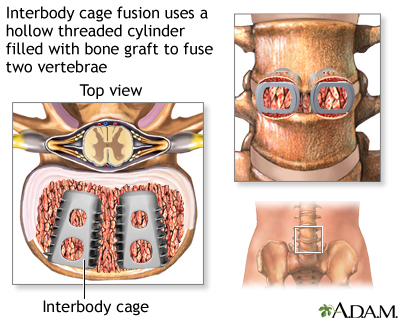
Interbody cage fusion
Interbody cage fusion uses a hollow threaded titanium or carbon fiber cylinder to fuse two vertebrae together. The diseased disk is removed and two interbody cages are placed in the opening where the diseased disk has been removed. The cages are filled with bone graft. The bone grows through the holes in the cages fusing the vertebrae.
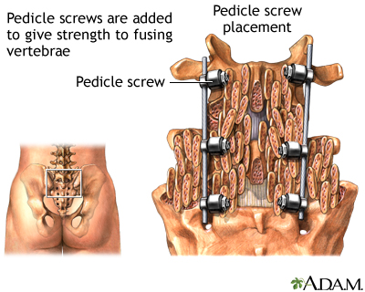
Pedicle screw
Pedicle screws are used sometimes in a spinal fusion to add extra support and strength to the fusion while it heals. Pedicle screws are placed above and below the vertebrae that were fused. A rod is used to connect the screws which prevents movement and allows the bone graft to heal. After the fusion is completely healed, the screws and rods can be removed. Removal isn't necessary unless they cause the patient discomfort.
Related Information
Spinal fusionBACK TO TOP
Review Date: 10/15/2023
Reviewed By: C. Benjamin Ma, MD, Professor, Chief, Sports Medicine and Shoulder Service, UCSF Department of Orthopaedic Surgery, San Francisco, CA. Also reviewed by David C. Dugdale, MD, Medical Director, Brenda Conaway, Editorial Director, and the A.D.A.M. Editorial team.

Health Content Provider
06/01/2025
|
A.D.A.M., Inc. is accredited by URAC, for Health Content Provider (www.urac.org). URAC's accreditation program is an independent audit to verify that A.D.A.M. follows rigorous standards of quality and accountability. A.D.A.M. is among the first to achieve this important distinction for online health information and services. Learn more about A.D.A.M.'s editorial policy, editorial process and privacy policy. A.D.A.M. is also a founding member of Hi-Ethics. This site complied with the HONcode standard for trustworthy health information from 1995 to 2022, after which HON (Health On the Net, a not-for-profit organization that promoted transparent and reliable health information online) was discontinued. |
The information provided herein should not be used during any medical emergency or for the diagnosis or treatment of any medical condition. A licensed medical professional should be consulted for diagnosis and treatment of any and all medical conditions. Links to other sites are provided for information only -- they do not constitute endorsements of those other sites. © 1997- 2025 A.D.A.M., a business unit of Ebix, Inc. Any duplication or distribution of the information contained herein is strictly prohibited.
 All rights reserved.