Chest tube insertion - series
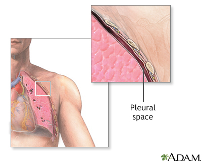
Normal anatomy
The pleural space is the space between the inner and outer lining of the lung. It is normally very thin, and lined only with a very small amount of fluid.
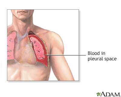
Indication
If fluid, such as blood, or air, gets into the pleural space, the lung can collapse, preventing adequate air exchange. Chest tubes are used to treat conditions that can cause the lung to collapse, such as:
- Air leaks from the lung into the chest (pneumothorax)
- Bleeding into the chest (hemothorax)
- After surgery or trauma in the chest (pneumothorax or hemothorax)
- Lung abscesses or pus in the chest (empyema)
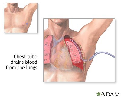
Procedure
Chest tubes are inserted to drain blood, fluid, or air and allow full expansion of the lungs. The tube is placed in the pleural space. The area where the tube will be inserted is numbed (local anesthesia). The patient may also be sedated. The chest tube is inserted between the ribs into the chest and is connected to a bottle or canister that contains sterile water. Suction is attached to the system to encourage drainage. A stitch (suture) and adhesive tape is used to keep the tube in place.
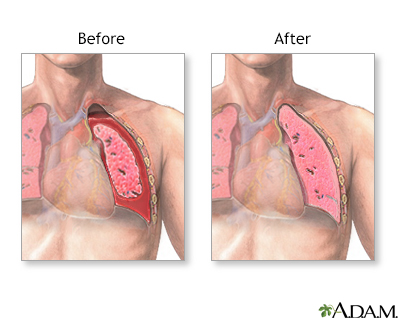
The chest tube usually remains in place until the X-rays show that all the blood, fluid, or air has drained from the chest and the lung has fully re-expanded. When the chest tube is no longer needed, it can be easily removed, usually without the need for medications to sedate or numb the patient. Medications may be used to prevent or treat infection (antibiotics).
Aftercare
Recovery from the chest tube insertion and removal is usually complete, with only a small scar.
The patient will stay in the hospital until the chest tube is removed. While the chest tube is in place, the nursing staff will carefully check for possible air leaks, breathing difficulties, and need for additional oxygen. Frequent deep breathing and coughing is necessary to help re-expand the lung, assist with drainage, and prevent normal fluids from collecting in the lungs.
Related Information
EmpyemaHemothorax
Collapsed lung (pneumothorax)
Chest tube insertion
BACK TO TOP
Review Date: 7/12/2024
Reviewed By: Jesse Borke, MD, CPE, FAAEM, FACEP, Attending Physician at Kaiser Permanente, Orange County, CA. Also reviewed by David C. Dugdale, MD, Medical Director, Brenda Conaway, Editorial Director, and the A.D.A.M. Editorial team.

Health Content Provider
06/01/2025
|
A.D.A.M., Inc. is accredited by URAC, for Health Content Provider (www.urac.org). URAC's accreditation program is an independent audit to verify that A.D.A.M. follows rigorous standards of quality and accountability. A.D.A.M. is among the first to achieve this important distinction for online health information and services. Learn more about A.D.A.M.'s editorial policy, editorial process and privacy policy. A.D.A.M. is also a founding member of Hi-Ethics. This site complied with the HONcode standard for trustworthy health information from 1995 to 2022, after which HON (Health On the Net, a not-for-profit organization that promoted transparent and reliable health information online) was discontinued. |
The information provided herein should not be used during any medical emergency or for the diagnosis or treatment of any medical condition. A licensed medical professional should be consulted for diagnosis and treatment of any and all medical conditions. Links to other sites are provided for information only -- they do not constitute endorsements of those other sites. © 1997- 2025 A.D.A.M., a business unit of Ebix, Inc. Any duplication or distribution of the information contained herein is strictly prohibited.
 All rights reserved.