CT angiography - head and neck
Computed tomography angiography - brain; CTA - skull; CTA - cranial; TIA-CTA head; Stroke-CTA head; Computed tomography angiography - neck; CTA - neck; Vertebral artery - CTA; Carotid artery stenosis - CTA; Vertebrobasilar - CTA; Posterior circulation ischemia - CTA; TIA - CTA neck; Stroke - CTA neck
CT angiography (CTA) combines a CT scan with the injection of dye. CT stands for computed tomography. This technique is able to create pictures of the blood vessels in the head and neck.
Images
How the Test is Performed
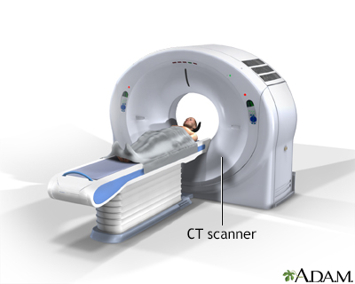
You will be asked to lie on a narrow table that slides into the center of the CT scanner.
While inside the scanner, the machine's x-ray beam rotates around you.
A computer creates many separate images of the body area, called slices. These images can be stored, viewed on a monitor, or printed on film. Three-dimensional models of the head and neck area can be created by stacking the slices together.
You must be still during the exam, because movement causes blurred images. You may be told to hold your breath for short periods of time.
Complete scans can usually be completed in a few minutes or less. The newest scanners can image your entire body, head to toe, in less than 30 seconds.
How to Prepare for the Test
Certain exams require a special dye, called contrast, to be delivered into the body before the test starts. Contrast helps certain areas show up better on x-rays.
- Contrast can be given through a vein (IV) in your hand or forearm. If contrast is used, you may also be asked not to eat or drink anything for 4 to 6 hours before the test.
- Let your health care provider know if you have ever had a reaction to contrast. You may need to take medicines before the test in order to safely receive it.
- Before receiving the contrast, tell your provider if you take the diabetes medicine metformin (Glucophage). You may need to take extra precautions.
The contrast can worsen kidney function problems in people with poorly functioning kidneys. Talk to your provider if you have a history of kidney problems.
Too much weight can damage the scanner. If you weigh more than 300 pounds (about 135 kilograms), talk to your provider about the weight limit before the test.
You will be asked to remove jewelry and wear a hospital gown during the study.
How the Test will Feel
Some people may have discomfort from lying on the hard table.
If you have contrast through a vein, you may have a:
- Slight burning feeling
- Metallic taste in your mouth
- Warm flushing of your body
This is normal and usually goes away within a few seconds.
Why the Test is Performed
CTA of the head may be done to look for the cause of:
- Changes in thinking or behavior
- Difficulty pronouncing words
- Dizziness or vertigo
- Double vision or vision loss
- Fainting
- Headache, when you have certain other signs or symptoms
- Hearing loss (in some people)
- Numbness or tingling, most often on the face or scalp
- Swallowing problems
- Stroke
- Bleeding in or around the brain (intracerebral hemorrhage, subarachnoid hemorrhage)
- Transient ischemic attack (TIA)
- Weakness in one part of your body
CTA of the neck may also be done:
- After trauma to the neck to look for damage to blood vessels
- For planning before carotid artery surgery
- For planning for brain tumor surgery
- For suspected vasculitis (inflammation of the blood vessel walls)
- For suspected abnormal blood vessels in the brain
Normal Results
Results are considered normal if no problems are seen.
What Abnormal Results Mean
Abnormal results may be due to:
- Abnormal blood vessels (arteriovenous malformation).
- Bleeding inside the brain or skull.
- Brain tumor or other growth (mass).
- Stroke.
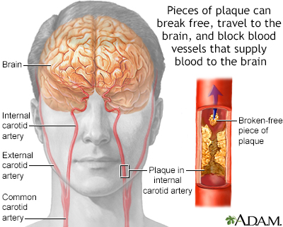
- Narrowed or blocked carotid arteries. (The carotid arteries provide the main blood supply to your brain. They are located on each side of your neck.)
- Narrowed or blocked vertebral artery in the neck. (The vertebral arteries provide blood flow to the back of the brain.)
- A tear in the wall of an artery (dissection).
- A weak area in the wall of a blood vessel that causes the blood vessel to bulge or balloon out (aneurysm).
Risks
Risks of CT scans include:
- Being exposed to radiation
- Allergic reaction to the contrast dye
- Damage to the kidneys from the dye
CT scans use more radiation than regular x-rays. Having many x-rays or CT scans over time may increase your risk for cancer. However, the risk from any one scan is small. You and your provider should weigh this risk against the benefits of getting a correct diagnosis for a medical problem. Most modern scanners use techniques to use less radiation.
Some people have allergies to contrast dye. Let your provider know if you have ever had an allergic reaction to injected contrast dye.
- The most common type of contrast given into a vein contains iodine. If you have an iodine allergy, you may have nausea or vomiting, sneezing, itching, or hives if you get this type of contrast.
- If you absolutely must be given such contrast, your provider may give you antihistamines (such as Benadryl) or steroids before the test.
- The kidneys help remove iodine out of the body. People with kidney disease or diabetes may need to receive extra fluids after the test to help flush the iodine out of the body.
Rarely, the dye may cause a life-threatening allergic response called anaphylaxis. Tell the scanner operator right away if you have any trouble breathing during the test. Scanners come with an intercom and speakers, so the operator can hear you at all times.
Considerations
A CT scan can reduce or avoid the need for invasive procedures to diagnose problems in the skull. This is one of the safest ways to study the head and neck.
Other tests that may be done instead of CT scan of the head include:
- MRI of the head
- Positron emission tomography (PET) scan of the head
- Conventional angiogram – a catheter injects dye directly into the vessels of the neck and brain
References
Barras CD, Bhattacharya JJ. Current status of imaging of the brain and anatomical features. In: Adam A, Dixon AK, Gillard JH, Schaefer-Prokop CM, eds. Grainger & Allison's Diagnostic Radiology: A Textbook of Medical Imaging. 7th ed. Philadelphia, PA: Elsevier; 2021:chap 53.
Wippold FJ, Orlowski HLP. Neuroradiology: the surrogate of gross neuropathology. In: Perry A, Brat DJ, eds. Practical Surgical Neuropathology: A Diagnostic Approach. 2nd ed. Philadelphia, PA: Elsevier; 2018:chap 4.
BACK TO TOPReview Date: 6/13/2024
Reviewed By: Joseph V. Campellone, MD, Department of Neurology, Cooper Medical School at Rowan University, Camden, NJ. Review provided by VeriMed Healthcare Network. Also reviewed by David C. Dugdale, MD, Medical Director, Brenda Conaway, Editorial Director, and the A.D.A.M. Editorial team.

Health Content Provider
06/01/2025
|
A.D.A.M., Inc. is accredited by URAC, for Health Content Provider (www.urac.org). URAC's accreditation program is an independent audit to verify that A.D.A.M. follows rigorous standards of quality and accountability. A.D.A.M. is among the first to achieve this important distinction for online health information and services. Learn more about A.D.A.M.'s editorial policy, editorial process and privacy policy. A.D.A.M. is also a founding member of Hi-Ethics. This site complied with the HONcode standard for trustworthy health information from 1995 to 2022, after which HON (Health On the Net, a not-for-profit organization that promoted transparent and reliable health information online) was discontinued. |
The information provided herein should not be used during any medical emergency or for the diagnosis or treatment of any medical condition. A licensed medical professional should be consulted for diagnosis and treatment of any and all medical conditions. Links to other sites are provided for information only -- they do not constitute endorsements of those other sites. © 1997- 2025 A.D.A.M., a business unit of Ebix, Inc. Any duplication or distribution of the information contained herein is strictly prohibited.
 All rights reserved.