Renal perfusion scintiscan
Renal perfusion scintigraphy; Radionuclide renal perfusion scan; Perfusion scintiscan - renal; Scintiscan - renal perfusion
A renal perfusion scintiscan is a nuclear medicine test. It uses a small amount of a radioactive substance to create an image of the kidneys.
Images
I Would Like to Learn About:
How the Test is Performed
You may be asked to take a blood pressure medicine called an ACE inhibitor, most often a medicine called captopril. The drug may be taken by mouth, or given through a vein (IV). The medicine makes the test more accurate.
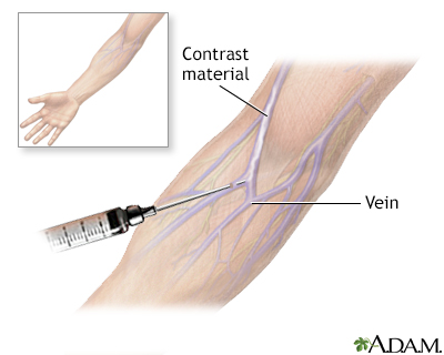
You will lie down on the scanner table shortly after taking the medicine. Your health care provider will inject a small amount of radioactive material (radioisotope) into one of your veins. Images of your kidneys are taken as the radioactive material flows through the arteries in the area. You will need to remain still for the entire test. The scan takes about 30 minutes.
About 10 minutes after you receive the radioactive material, you will be given a diuretic ("water pill") through a vein. This medicine also helps make the test more accurate.
You can return to normal activities right after the test. You should drink plenty of fluids to help remove the radioactive material from your body. The test will make you urinate more frequently for several hours after the test.
How to Prepare for the Test
You will be asked to drink plenty of water before the test.
If you are currently taking an ACE inhibitor for high blood pressure, you may be asked to stop taking your medicine before the exam. Always talk to your provider before you stop any of your medicines.
You may be asked to wear a hospital gown. Remove all jewelry and metallic objects before the scan.
How the Test will Feel
You may feel a small amount of pain when the needle is inserted.
You must remain still during the scan. You will be told when you need to change positions.
There may be some discomfort as your bladder fills with urine during the exam. Tell the person conducting the exam if you must urinate before the scan is complete.
Why the Test is Performed
The test evaluates blood flow to the kidneys. It is used to diagnose narrowing of the arteries that supply the kidneys. This is a condition called renal artery stenosis. Significant renal artery stenosis may be a cause of high blood pressure and kidney problems.
Normal Results
Blood flow to the kidneys appears normal.
What Abnormal Results Mean
Abnormal findings on the scan may be a sign of renal artery stenosis. A similar test that does not use an ACE inhibitor can be done to confirm the diagnosis.
Risks
If you are pregnant or nursing, your provider may want to postpone the test. Pregnant women should not take ACE inhibitors.
The amount of radioactivity in the injection is very small. Nearly all radioactivity is gone from the body within 24 hours.
Reactions to the materials used during this test are rare, but may include rash, swelling, or anaphylaxis.
Risks of a needle stick are slight, but include infection and bleeding.
Considerations
This test may be less accurate in people who already have kidney disease. Talk to your provider to determine if this is the right test for you. Alternatives to this test are an MRI or CT angiogram.
References
Duddalwar VA, Jadvar H, Palmer SL. Diagnostic kidney imaging. In: Yu ASL, Chertow GM, Luyckx VA, Marsden PA, Skorecki K, Taal MW, eds. Brenner and Rector's The Kidney. 11th ed. Philadelphia, PA: Elsevier; 2020:chap 25.
Rottenberg G, Lagha E. Renal transplantation: imaging. In: Adam A, Dixon AK, Gillard JH, Schaefer-Prokop CM, eds. Grainger & Allison's Diagnostic Radiology. 7th ed. Philadelphia, PA: Elsevier; 2021:chap 30.
Textor SC. Renovascular hypertension and ischemic nephropathy. In: Yu ASL, Chertow GM, Luyckx VA, Marsden PA, Skorecki K, Taal MW, eds. Brenner and Rector's The Kidney. 11th ed. Philadelphia, PA: Elsevier; 2020:chap 47.
BACK TO TOPReview Date: 1/1/2025
Reviewed By: Kelly L. Stratton, MD, FACS, Associate Professor, Department of Urology, University of Oklahoma Health Sciences Center, Oklahoma City, OK. Also reviewed by David C. Dugdale, MD, Medical Director, Brenda Conaway, Editorial Director, and the A.D.A.M. Editorial team.

Health Content Provider
06/01/2025
|
A.D.A.M., Inc. is accredited by URAC, for Health Content Provider (www.urac.org). URAC's accreditation program is an independent audit to verify that A.D.A.M. follows rigorous standards of quality and accountability. A.D.A.M. is among the first to achieve this important distinction for online health information and services. Learn more about A.D.A.M.'s editorial policy, editorial process and privacy policy. A.D.A.M. is also a founding member of Hi-Ethics. This site complied with the HONcode standard for trustworthy health information from 1995 to 2022, after which HON (Health On the Net, a not-for-profit organization that promoted transparent and reliable health information online) was discontinued. |
The information provided herein should not be used during any medical emergency or for the diagnosis or treatment of any medical condition. A licensed medical professional should be consulted for diagnosis and treatment of any and all medical conditions. Links to other sites are provided for information only -- they do not constitute endorsements of those other sites. © 1997- 2025 A.D.A.M., a business unit of Ebix, Inc. Any duplication or distribution of the information contained herein is strictly prohibited.
 All rights reserved.