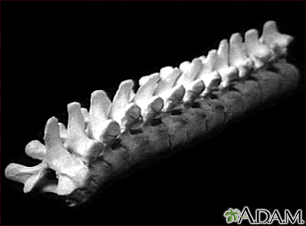
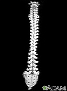
Thoracic spine x-ray
Vertebral radiography; X-ray - spine; Thoracic x-ray; Spine x-ray; Thoracic spine films; Back films
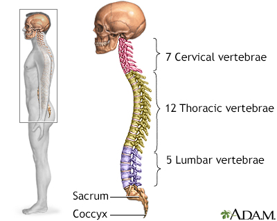
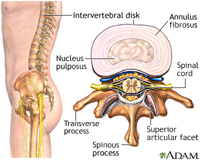
A thoracic spine x-ray is an x-ray of the 12 chest (thoracic) bones (vertebrae) of the spine. The vertebrae are separated by flat pads of cartilage called disks that provide a cushion between the bones.
Images
I Would Like to Learn About:
How the Test is Performed
The test is done in a hospital radiology department or in your health care provider's office. You will lie on the x-ray table in different positions. If the x-ray is checking for an injury, care will be taken to prevent further injury.
The x-ray machine will be moved over the thoracic area of the spine. You will hold your breath as the picture is taken so that the picture will not be blurry. Usually, 2 or 3 x-ray views are needed.
How to Prepare for the Test
Tell your provider if you are pregnant. Also tell your provider if you have had surgery in your chest, abdomen, or pelvis.
Remove all jewelry.
How the Test will Feel
The test causes no discomfort. The table may be cold.
Why the Test is Performed
The x-ray helps evaluate:
- Bone injuries
- Cartilage loss
- Arthritis
- Curvature in the spine
- Diseases of the bone
- Tumors of the bone
What Abnormal Results Mean
The test can detect:
- Bone spurs
- Deformities of the spine
- Disk narrowing
- Dislocations
- Fractures (most often compression fractures of the vertebrae)
- Thinning of the bone (osteoporosis)
- Wearing away (degeneration) of the vertebrae
- Abnormal alignment of the bone (spondylolisthesis)
Risks
There is low radiation exposure. X-rays are monitored and regulated to provide the minimum amount of radiation exposure needed to produce the image. Most experts feel that the risk is low compared with the benefits.
Pregnant women and children are more sensitive to the risks of x-rays.
Considerations
The x-ray will not detect problems in the muscles, nerves, and other soft tissues, because these problems cannot be seen well on an x-ray.
Related Information
X-rayBroken bone
Osteoporosis
References
Preston-Suni K, Kaji AH. Spinal trauma. In: Walls RM, ed. Rosen's Emergency Medicine: Concepts and Clinical Practice. 10th ed. Philadelphia, PA: Elsevier; 2023:chap 35.
Mettler FA. Skeletal system. In: Mettler FA, ed. Essentials of Radiology. 4th ed. Philadelphia, PA: Elsevier; 2019:chap 8.
Van Thielen T, van den Hauwe L, Van Goethem JW, Parizel PM. Current status of imaging of the spine and anatomical features. In: Adam A, Dixon AK, Gillard JH, Schaefer-Prokop CM, eds. Grainger & Allison's Diagnostic Radiology: A Textbook of Medical Imaging. 7th ed. Philadelphia, PA: Elsevier; 2021:chap 47.
BACK TO TOPReview Date: 8/12/2023
Reviewed By: C. Benjamin Ma, MD, Professor, Chief, Sports Medicine and Shoulder Service, UCSF Department of Orthopaedic Surgery, San Francisco, CA. Also reviewed by David C. Dugdale, MD, Medical Director, Brenda Conaway, Editorial Director, and the A.D.A.M. Editorial team.

Health Content Provider
06/01/2025
|
A.D.A.M., Inc. is accredited by URAC, for Health Content Provider (www.urac.org). URAC's accreditation program is an independent audit to verify that A.D.A.M. follows rigorous standards of quality and accountability. A.D.A.M. is among the first to achieve this important distinction for online health information and services. Learn more about A.D.A.M.'s editorial policy, editorial process and privacy policy. A.D.A.M. is also a founding member of Hi-Ethics. This site complied with the HONcode standard for trustworthy health information from 1995 to 2022, after which HON (Health On the Net, a not-for-profit organization that promoted transparent and reliable health information online) was discontinued. |
The information provided herein should not be used during any medical emergency or for the diagnosis or treatment of any medical condition. A licensed medical professional should be consulted for diagnosis and treatment of any and all medical conditions. Links to other sites are provided for information only -- they do not constitute endorsements of those other sites. © 1997- 2025 A.D.A.M., a business unit of Ebix, Inc. Any duplication or distribution of the information contained herein is strictly prohibited.
 All rights reserved.