Head MRI
Nuclear magnetic resonance - cranial; Magnetic resonance imaging - cranial; MRI of the head; MRI - cranial; NMR - cranial; Cranial MRI; Brain MRI; MRI - brain; MRI - head
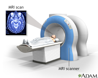
A head MRI (magnetic resonance imaging) is an imaging test that uses powerful magnets and radio waves to create pictures of the brain and surrounding tissues.
It does not use radiation.
Images
How the Test is Performed
A head MRI is done in the hospital or a radiology center.
You lie on a narrow table, which slides into a large tunnel-shaped scanner.
Some MRI exams require a special dye, called contrast material. The dye is usually given during the test through a vein (IV) in your hand or forearm. The dye helps the radiologist see certain areas more clearly.
During the MRI, the person who operates the machine watches you from another room. The test most often lasts 30 to 60 minutes, but may take longer.
How to Prepare for the Test
You may be asked not to eat or drink anything for 4 to 6 hours before the scan.
Tell your health care provider if you are afraid of close spaces (have claustrophobia). You may receive medicine to help you feel sleepy and less anxious. Or your provider may suggest an open MRI, in which the machine is not as close to the body.
You may be asked to wear a hospital gown or clothing without metal ties (such as sweatpants and a t-shirt). Certain types of metal can cause blurry images.
Before the test, tell your provider if you have:
- Brain aneurysm clips
- An artificial heart valve
- Heart defibrillator or pacemaker
- Inner ear (cochlear) implants
- Kidney disease or are on dialysis (you may not be able to receive contrast)
- Recently placed artificial joint
- A blood vessel stent
- Worked with sheet metal in the past (you may need tests to check for metal pieces in your eyes)
The MRI contains strong magnets. Metal objects are not allowed into the room with the MRI scanner. This includes:
- Pens, pocketknives, and eyeglasses
- Items such as jewelry, watches, credit cards, and hearing aids
- Pins, hairpins, metal zippers, and similar metallic items
- Removable dental work
How the Test will Feel
If you need dye, you will feel the needle pinch in your arm when the dye is injected into the vein.
An MRI exam causes no pain. If you have difficulty lying still or are very nervous, you may be given a medicine to relax. Too much movement can blur the images and cause errors.
The table may be hard or cold, but you can ask for a blanket or pillow. The machine makes loud thumping and humming noises when turned on. You can ask for ear plugs to help reduce the noise.
An intercom in the room allows you to speak to someone at any time. Some MRIs have televisions and special headphones that can help you pass the time or block the scanner noise.
There is no recovery time, unless you were given a medicine to relax. After an MRI scan, you can go back to your normal diet, activity, and medicines.
Why the Test is Performed
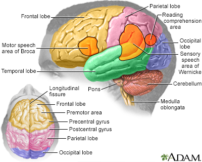
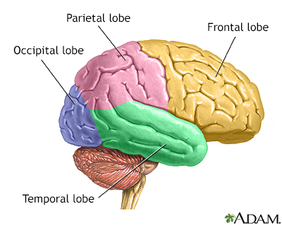
An MRI provides detailed pictures of the brain and nerve tissues.
A brain MRI can be used to diagnose and monitor many diseases and disorders that affect the brain, including:
- Birth defect
- Bleeding (subarachnoid, subdural or epidural bleed, or bleeding in the brain tissue itself)
- Aneurysms
- Infection, such as brain abscess
- Tumors (cancerous and noncancerous)
- Hormonal disorders (such as acromegaly, galactorrhea, and Cushing syndrome)
- Multiple sclerosis
- Stroke
An MRI scan of the head can also determine the cause of:
- Muscle weakness or numbness and tingling
- Changes in thinking or behavior
- Hearing loss
- Headaches when certain other symptoms or signs are present
- Speaking difficulties
- Vision problems
- Dementia
A special type of MRI called magnetic resonance angiography (MRA) may be done to look at blood vessels in the brain.
What Abnormal Results Mean
Abnormal results may be due to:
- Abnormal blood vessels in the brain (arteriovenous malformations of the head)
- Tumor of the nerve that connects the ear to the brain (acoustic neuroma)
- Bleeding in or around the brain
- Brain infection
- Brain tissue swelling
- Brain tumors
- Damage to the brain from an injury
- Fluid collecting around the brain (hydrocephalus)
- Infection of the skull bones (osteomyelitis)
- Loss of brain tissue
- Multiple sclerosis
- Stroke or transient ischemic attack (TIA)
- Structural problems in the brain
Risks
MRI uses no radiation. To date, no side effects from the magnetic fields and radio waves have been reported.
The most common type of contrast (dye) used is gadolinium. It is very safe. Allergic reactions to this substance rarely occur. However, gadolinium can be harmful to people with kidney problems who are on dialysis. If you have kidney problems, tell your provider before the test.
The strong magnetic fields created during an MRI can make heart pacemakers and other implants not work as well. It can also cause a piece of metal inside your body to move or shift.
Considerations
MRI is safe during pregnancy. In many cases MRI may be more sensitive than CT scan to problems in the brain such as small masses. CT is usually better at looking for small areas of bleeding.
Tests that may be done instead of an MRI of the head include:
- Head CT scan
- Positron emission tomography (PET) scan of the brain
A CT scan may be preferred in the following cases, since it is faster and usually available right in the emergency room:
- Acute trauma of the head and face
- Bleeding in the brain (within the first 24 to 48 hours)
- Early symptoms of stroke
- Skull bone disorders and disorders involving the bones of the ear
Related Information
MRICT scan
Multiple sclerosis
Myelin
Arteriogram
Brain tumor - children
Metastatic brain tumor
Pituitary tumor
Acoustic neuroma
Optic glioma
Cerebral arteriovenous malformation
Aneurysm in the brain
Subdural hematoma
Blood clots
Osmotic demyelination syndrome
Brain abscess
Amyotrophic lateral sclerosis (ALS)
Chronic subdural hematoma
Dementia
Dementia due to metabolic causes
Central diabetes insipidus
Huntington disease
Hypopituitarism
Melanoma of the eye
Meniere disease
Vascular dementia
Normal pressure hydrocephalus
Focal seizure
Absence seizure
Cushing disease
Prolactinoma
Reye syndrome
Cerebral amyloid angiopathy
Alzheimer disease
Sinusitis
Stroke
TMJ disorders
Toxoplasmosis
Wernicke-Korsakoff syndrome
Wilson disease
References
Barras CD, Bhattacharya JJ. Current status of imaging of the brain and anatomical features. In: Adam A, Dixon AK, Gillard JH, Schaefer-Prokop CM, eds. Grainger & Allison's Diagnostic Radiology. 7th ed. Philadelphia, PA: Elsevier; 2021:chap 53.
Kalnins AU, Prost RW, Kim TA. Computed tomography and magnetic resonance imaging of the brain. In: Winn HR, eds. Youmans and Winn Neurological Surgery. 8th ed. Philadelphia, PA: Elsevier; 2023:chap 10.
Khan M, Schulte J, Zinreich SJ, Aygun N. Overview of diagnostic imaging of the head and neck. In: Flint PW, Francis HW, Haughey BH, et al, eds. Cummings Otolaryngology: Head and Neck Surgery. 7th ed. Philadelphia, PA: Elsevier; 2021:chap 8.
BACK TO TOPReview Date: 7/15/2024
Reviewed By: Jason Levy, MD, FSIR, Northside Radiology Associates, Atlanta, GA. Also reviewed by David C. Dugdale, MD, Medical Director, Brenda Conaway, Editorial Director, and the A.D.A.M. Editorial team.

Health Content Provider
06/01/2025
|
A.D.A.M., Inc. is accredited by URAC, for Health Content Provider (www.urac.org). URAC's accreditation program is an independent audit to verify that A.D.A.M. follows rigorous standards of quality and accountability. A.D.A.M. is among the first to achieve this important distinction for online health information and services. Learn more about A.D.A.M.'s editorial policy, editorial process and privacy policy. A.D.A.M. is also a founding member of Hi-Ethics. This site complied with the HONcode standard for trustworthy health information from 1995 to 2022, after which HON (Health On the Net, a not-for-profit organization that promoted transparent and reliable health information online) was discontinued. |
The information provided herein should not be used during any medical emergency or for the diagnosis or treatment of any medical condition. A licensed medical professional should be consulted for diagnosis and treatment of any and all medical conditions. Links to other sites are provided for information only -- they do not constitute endorsements of those other sites. © 1997- 2025 A.D.A.M., a business unit of Ebix, Inc. Any duplication or distribution of the information contained herein is strictly prohibited.
 All rights reserved.