Orbit CT scan
CT scan - orbital; Eye CT scan; Computed tomography scan - orbit
A computed tomography (CT) scan of the orbit is an imaging method. It uses x-rays to create detailed pictures of the eye sockets (orbits), eyes and surrounding bones.
Images
I Would Like to Learn About:
How the Test is Performed
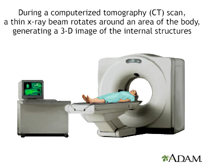
You will be asked to lie on a narrow table that slides into the center of the CT scanner. Only your head is placed inside the CT scanner.
You may lie on your back, or you may lie face-down with your chin raised. You may be allowed to rest your head on a pillow.
Once you are inside the scanner, the machine's x-ray beam rotates around you, but you won't see the x-ray.
A computer creates separate images of the body area, called slices. These images can be stored, viewed on a monitor, or printed on film. The computer can create three-dimensional models of the body area by stacking the slices together.
You must lie still during the exam, because movement causes blurred images. You may be asked to hold your breath for short periods. You may be asked to remove jewelry and earrings.
The actual scan takes about 30 seconds. The entire process takes about 15 minutes.
How to Prepare for the Test
Before the test:
- You will be asked to remove jewelry and wear a hospital gown during the study.
- If you weigh more than 300 pounds (135 kilograms), find out if the CT machine has a weight limit. Too much weight can cause damage to the scanner's working parts or injury to the person.
Certain exams require a special dye, called contrast, to be delivered into the body before the test starts. Contrast helps certain areas show up better on the x-rays. Contrast can be given through a vein (intravenous - IV) in your hand or forearm.
Before a scan using contrast, it is important to know the following:
- You may be asked not to eat or drink anything for 4 to 6 hours before the test.
- Let your health care provider know if you have ever had a reaction to contrast. You may need to take medicines before the test in order to safely receive this substance.
- Tell your provider if you take the diabetes medicine metformin (Glucophage). You may need to take extra precautions.
- Let your provider know if you have poor kidney function. This is because the contrast can worsen kidney function.
How the Test will Feel
Some people may have discomfort from lying on the hard table.
Contrast given through an IV may cause a slight burning sensation. You may also have a metallic taste in the mouth and a warm flushing of the body. These sensations are normal and most often go away within a few seconds.
Why the Test is Performed
This test is helpful for diagnosing diseases that affect the following areas around the eyes:
- Blood vessels
- Eye muscles
- Nerves supplying the eyes (optic nerves)
- Sinuses
An orbit CT scan may also be used to detect:
- Abscess (infection) of the eye area
- Broken eye socket bone
- Foreign object in the eye socket
What Abnormal Results Mean
Abnormal results may mean:
- Bleeding
- Broken eye socket bone
- Graves disease
- Infection
- Tumor
Risks
CT scans and other x-rays are strictly monitored and controlled to make sure they use the least amount of radiation. The risk associated with any individual scan is very low. The risk increases as more scans are done.
CT scans are done when the benefits greatly outweigh the risks. For example, it can be more risky not to have the exam, especially if your provider thinks you might have cancer or a fracture.
The most common type of contrast given into a vein contains iodine.
- If a person with an iodine allergy is given this type of contrast, nausea, sneezing, vomiting, itching, or hives may occur.
- If you have a known allergy to contrast but need it for a successful exam, you may receive antihistamines (such as Benadryl) or steroids before the test.
The kidneys help filter the iodine out of the body. If you have kidney disease or diabetes, you should be closely monitored for kidney problems after contrast is given. If you have kidney disease or diabetes, talk to your provider before the test to know your risks.
Before receiving the contrast, tell your provider if you take the diabetes medicine metformin (Glucophage) because you may need to take extra precautions. You may need to stop the medicine for 48 hours after the test.
In rare cases, the dye can cause a life-threatening allergic response called anaphylaxis. If you have any trouble breathing during the test, tell the scanner operator right away. Scanners come with an intercom and speakers, so the operator can hear you at all times.
Related Information
CT scanGraves disease
Tumor
References
Campion T, Miszkiel K, Davagnanam I. The orbit. In: Adam A, Dixon AK, Gillard JH, Schaefer-Prokop CM, eds. Grainger & Allison's Diagnostic Radiology. 7th ed. Philadelphia, PA: Elsevier; 2021:chap 60.
Guluma K, Lee JE. Ophthalmology. In: Walls RM, ed. Rosen's Emergency Medicine: Concepts and Clinical Practice. 10th ed. Philadelphia, PA: Elsevier; 2023:chap 57.
Poon CS, Abrahams M, Abrahams JJ. Orbit. In: Haaga JR, Boll DT, eds. CT and MRI of the Whole Body. 6th ed. Philadelphia, PA: Elsevier; 2017:chap 20.
Salmon JF. Orbit. In: Salmon JF, ed. Kanski's Clinical Ophthalmology. 10th ed. Philadelphia, PA: Elsevier; 2025:chap 4.
BACK TO TOPReview Date: 1/1/2025
Reviewed By: Jason Levy, MD, FSIR, Northside Radiology Associates, Atlanta, GA. Also reviewed by David C. Dugdale, MD, Medical Director, Brenda Conaway, Editorial Director, and the A.D.A.M. Editorial team.

Health Content Provider
06/01/2025
|
A.D.A.M., Inc. is accredited by URAC, for Health Content Provider (www.urac.org). URAC's accreditation program is an independent audit to verify that A.D.A.M. follows rigorous standards of quality and accountability. A.D.A.M. is among the first to achieve this important distinction for online health information and services. Learn more about A.D.A.M.'s editorial policy, editorial process and privacy policy. A.D.A.M. is also a founding member of Hi-Ethics. This site complied with the HONcode standard for trustworthy health information from 1995 to 2022, after which HON (Health On the Net, a not-for-profit organization that promoted transparent and reliable health information online) was discontinued. |
The information provided herein should not be used during any medical emergency or for the diagnosis or treatment of any medical condition. A licensed medical professional should be consulted for diagnosis and treatment of any and all medical conditions. Links to other sites are provided for information only -- they do not constitute endorsements of those other sites. © 1997- 2025 A.D.A.M., a business unit of Ebix, Inc. Any duplication or distribution of the information contained herein is strictly prohibited.
 All rights reserved.