Intravenous pyelogram
Excretory urography; IVP
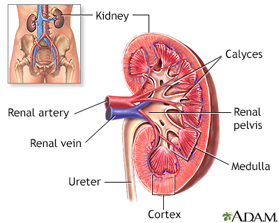
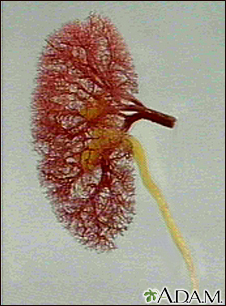
An intravenous pyelogram (IVP) is a special x-ray exam of the kidneys, bladder, and ureters (the tubes that carry urine from the kidneys to the bladder).
Images
I Would Like to Learn About:
How the Test is Performed
An IVP is done in a hospital radiology department or a health care provider's office.
You may be asked to take some medicine to clear your bowels before the procedure to provide a better view of the urinary tract. You will need to empty your bladder right before the procedure starts.
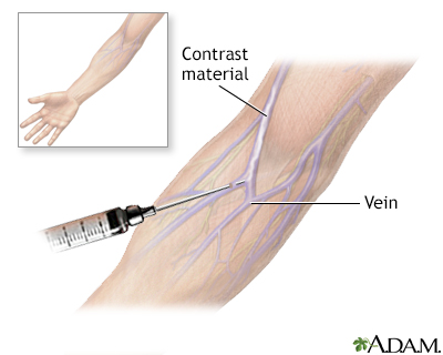
Your provider will inject an iodine-based contrast (dye) into a vein in your arm. A series of x-ray images are taken at different times. This is to see how the kidneys remove the dye and how it collects in your urine.
You will need to lie still during the procedure. The test may take up to an hour.
Before the final image is taken, you will be asked to urinate again. This is to see how well the bladder has emptied.
You can go back to your normal diet and medicines after the procedure. You should drink plenty of fluids to help remove all the contrast dye from your body.
How to Prepare for the Test
As with all x-ray procedures, tell your provider if you:
- Are allergic to contrast material
- Are pregnant
- Have any medicine allergies
- Have kidney disease or diabetes
Your provider will tell you if you can eat or drink before this test. You may be given a laxative to take the afternoon before the procedure to clear the intestines. This will help your kidneys to be seen clearly.
You must sign a consent form. You will be asked to wear a hospital gown and to remove all jewelry.
How the Test will Feel
You may feel a burning or flushing sensation in your arm and body as the contrast dye is injected. You may also have a metallic taste in your mouth. This is normal and will go away quickly.
Some people develop a headache, nausea, or vomiting after the dye is injected.
The belt across the kidneys may feel tight over your belly area.
Why the Test is Performed
An IVP can be used to evaluate:
- An abdominal injury
- Bladder and kidney infections
- Blood in the urine
- Flank pain (possibly due to kidney stones)
- Tumors
What Abnormal Results Mean
The test may reveal kidney diseases, birth defects of the urinary system, tumors, kidney stones, or damage to the urinary system.
Risks
There is a chance of an allergic reaction to the dye, even if you have received contrast dye in the past without any problem. If you have a known allergy to iodine-based contrast, a different test can be done. Other tests include retrograde pyelography, magnetic resonance imaging (MRI), or ultrasound.
There is low radiation exposure. Most experts feel that the risk is low compared with the benefits.
Children are more sensitive to the risks of radiation. This test is generally avoided during pregnancy.
Considerations
Computed tomography (CT) scans have replaced IVP as the main tool for checking the urinary system. MRI is also used to look at the kidneys, ureters, and bladder.
Related Information
X-rayUrine - bloody
Flank pain
Kidney stones
Acute arterial occlusion - kidney
Analgesic nephropathy
Atheroembolic renal disease
Enlarged prostate
Bilateral hydronephrosis
Renal pelvis or ureter cancer
Cystinuria
Injury - kidney and ureter
Autosomal dominant tubulointerstitial kidney disease
Polycystic kidney disease
Prostate cancer
Renal cell carcinoma
Renovascular hypertension
Retroperitoneal fibrosis
Ureterocele
Wilms tumor
References
Bishoff JT, Rastinehad AR. Urinary tract imaging: basic principles of CT, MRI, and plain film imaging. In: Partin AW, Dmochowski RR, Kavoussi LR, Peters CA, eds. Campbell-Walsh-Wein Urology. 12th ed. Philadelphia, PA: Elsevier; 2021:chap 3.
Gallagher KM, Hughes J. Urinary tract obstruction. In: Johnson RJ, Floege J, Tonelli M, eds. Comprehensive Clinical Nephrology. 7th ed. Philadelphia, PA: Elsevier; 2024:chap 61.
Sakhaee K, Moe OW. Urolithiasis. In: Yu ASL, Chertow GM, Luyckx VA, Marsden PA, Skorecki K, Taal MW, eds. Brenner and Rector's The Kidney. 11th ed. Philadelphia, PA: Elsevier; 2020:chap 38.
BACK TO TOPReview Date: 1/1/2025
Reviewed By: Kelly L. Stratton, MD, FACS, Associate Professor, Department of Urology, University of Oklahoma Health Sciences Center, Oklahoma City, OK. Also reviewed by David C. Dugdale, MD, Medical Director, Brenda Conaway, Editorial Director, and the A.D.A.M. Editorial team.

Health Content Provider
06/01/2025
|
A.D.A.M., Inc. is accredited by URAC, for Health Content Provider (www.urac.org). URAC's accreditation program is an independent audit to verify that A.D.A.M. follows rigorous standards of quality and accountability. A.D.A.M. is among the first to achieve this important distinction for online health information and services. Learn more about A.D.A.M.'s editorial policy, editorial process and privacy policy. A.D.A.M. is also a founding member of Hi-Ethics. This site complied with the HONcode standard for trustworthy health information from 1995 to 2022, after which HON (Health On the Net, a not-for-profit organization that promoted transparent and reliable health information online) was discontinued. |
The information provided herein should not be used during any medical emergency or for the diagnosis or treatment of any medical condition. A licensed medical professional should be consulted for diagnosis and treatment of any and all medical conditions. Links to other sites are provided for information only -- they do not constitute endorsements of those other sites. © 1997- 2025 A.D.A.M., a business unit of Ebix, Inc. Any duplication or distribution of the information contained herein is strictly prohibited.
 All rights reserved.