Renal arteriography
Renal angiogram; Angiography - kidney; Renal angiography; Renal artery stenosis - arteriography
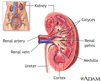
Renal arteriography is a special x-ray of the arteries of the kidneys.
Images
I Would Like to Learn About:
How the Test is Performed
This test is done in the hospital or outpatient office by a radiologist usually assisted by a technician or other staff members. You will lie on an x-ray table.
Doctors often use an artery near the groin for the test. Occasionally, the doctor may use an artery in the wrist.
Your doctor will:
- Clean and shave the area.
- Apply a numbing medicine to the area.
- Place a needle into the artery.
- Pass a thin wire through the needle into the artery.
- Take out the needle.
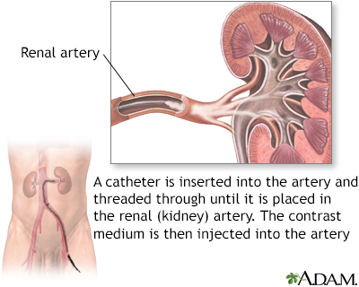
- Insert a long, narrow, flexible tube called a catheter in its place.
The doctor directs the catheter into correct position using x-ray images of the body. An instrument called fluoroscope sends the images to a TV monitor, which the doctor can see.
The catheter is pushed ahead over the wire into the aorta (main blood vessel from the heart). It then enters the kidney artery. The test uses a special dye (called contrast) to help the arteries show on the x-ray. The blood vessels of the kidneys are not seen with ordinary x-rays. The dye flows through the catheter into the kidney artery.
X-ray images are taken as the dye moves through the blood vessels. Saline (sterile salt water) containing a blood thinner may also be sent through the catheter to keep blood in the area from clotting.
The catheter is removed after the x-rays are taken. A closure device is placed in the groin or pressure is applied to the area to stop the bleeding. You may be asked to keep your leg straight for 4 to 6 hours after the procedure.
How to Prepare for the Test
Tell your doctor if:
- You are pregnant
- You have ever had any bleeding problems
- You currently take blood thinners, including daily aspirin
- You ever had any allergic reactions, especially those related to x-ray contrast material or iodine substances
- You have ever been diagnosed with kidney failure or poorly functioning kidneys
You must sign a consent form. DO NOT eat or drink anything for 6 to 8 hours before the test. You will be given a hospital gown to wear and asked to remove all jewelry. You may be given a pain pill (sedative) before the procedure or IV sedatives during the procedure.
How the Test will Feel
You will lie flat on the x-ray table. There is usually a cushion, but it is not as comfortable as a bed. You may feel a sting when the anesthesia medicine is given. You may feel some pressure and discomfort as the catheter is positioned.
Some people feel a warm sensation when the dye is injected, but most people cannot feel it. You don't feel the catheter inside your body.
There may be slight tenderness and bruising at the site of the injection after the test.
Why the Test is Performed
Renal arteriography is often done after other tests are done first. These may include duplex ultrasound, CT abdomen, CT angiogram, MRI abdomen, or MRI angiogram. These tests may show the following problems.
- Abnormal widening of an artery, called an aneurysm
- Abnormal connections between veins and arteries (fistulas)
- Blood clot blocking an artery supplying the kidney
- Unexplained high blood pressure thought to be due to narrowing of the blood vessels of the kidneys
- Benign tumors and cancers involving the kidneys
- Active bleeding from the kidney
If these tests show problems renal arteriography may be used to guide a treatment such as embolization or arterial stent placement.
Normal Results
Results may vary. Talk to your health care provider about the meaning of your specific test results.
What Abnormal Results Mean
Renal angiography may show the presence of tumors, narrowing of the artery or aneurysms (widening of the vein or artery), blood clots, fistulas, or bleeding in the kidney.
The test may also be done with the following conditions:
- Blockage of an artery by a blood clot
- Renal artery stenosis
- Renal cell cancer
- Angiomyolipomas (noncancerous tumors of the kidney)
Some of these problems can be treated with techniques done at the same time the arteriogram is performed.
- Angioplasty is a procedure to open a narrowed or blocked blood vessels that supply blood to your kidneys.
- A stent is a small, metal mesh tube that keeps the artery open. It may be placed to keep a narrowed artery open.
- Cancers and noncancerous tumors can be treated using a process called embolization. This involves using substances that block blood flow in order to kill or shrink the tumor. Sometimes, this is performed in combination with surgery.
- Bleeding can also be treated with embolization.
Risks
The procedure is generally safe. There may be some risks, such as:
- Allergic reaction to the dye (contrast medium)
- Arterial damage
- Damage to the artery or artery wall, which can lead to blood clots
- Kidney damage from damage to the artery or from the dye
There is low radiation exposure. Pregnant women and children are more sensitive to the risks related to x-rays.
Considerations
The test should NOT be done if you are pregnant or have severe bleeding problems.
Magnetic resonance angiography (MRA) or CT angiography (CTA) can be done instead. MRA and CTA are noninvasive and can provide similar imaging of the kidney arteries, although they cannot be used for treatment.
Related Information
Renal venogramX-ray
Blood clots
Aneurysm
Acute arterial occlusion - kidney
Acute kidney failure
Atheroembolic renal disease
Renal cell carcinoma
References
Azarbal AF, Mclafferty RB. Arteriography. In: Sidawy AN, Perler BA, eds. Rutherford's Vascular Surgery and Endovascular Therapy. 10th ed. Philadelphia, PA: Elsevier; 2023:chap 27.
Duddalwar VA, Jadvar H, Palmer SL. Diagnostic kidney imaging. In: Yu ASL, Chertow GM, Luyckx VA, Marsden PA, Skorecki K, Taal MW, eds. Brenner and Rector's The Kidney. 11th ed. Philadelphia, PA: Elsevier; 2020:chap 25.
Textor SC. Renovascular hypertension and ischemic nephropathy. In: Yu ASL, Chertow GM, Luyckx VA, Marsden PA, Skorecki K, Taal MW, eds. Brenner and Rector's The Kidney. 11th ed. Philadelphia, PA: Elsevier; 2020:chap 47.
BACK TO TOPReview Date: 1/29/2024
Reviewed By: Jason Levy, MD, FSIR, Northside Radiology Associates, Atlanta, GA. Also reviewed by David C. Dugdale, MD, Medical Director, Brenda Conaway, Editorial Director, and the A.D.A.M. Editorial team.

Health Content Provider
06/01/2025
|
A.D.A.M., Inc. is accredited by URAC, for Health Content Provider (www.urac.org). URAC's accreditation program is an independent audit to verify that A.D.A.M. follows rigorous standards of quality and accountability. A.D.A.M. is among the first to achieve this important distinction for online health information and services. Learn more about A.D.A.M.'s editorial policy, editorial process and privacy policy. A.D.A.M. is also a founding member of Hi-Ethics. This site complied with the HONcode standard for trustworthy health information from 1995 to 2022, after which HON (Health On the Net, a not-for-profit organization that promoted transparent and reliable health information online) was discontinued. |
The information provided herein should not be used during any medical emergency or for the diagnosis or treatment of any medical condition. A licensed medical professional should be consulted for diagnosis and treatment of any and all medical conditions. Links to other sites are provided for information only -- they do not constitute endorsements of those other sites. © 1997- 2025 A.D.A.M., a business unit of Ebix, Inc. Any duplication or distribution of the information contained herein is strictly prohibited.
 All rights reserved.