Carotid duplex
Scan - carotid duplex; Carotid ultrasound; Carotid artery ultrasound; Ultrasound - carotid; Vascular ultrasound - carotid; Ultrasound - vascular - carotid; Stroke - carotid duplex; TIA - carotid duplex; Transient ischemic attack - carotid duplex
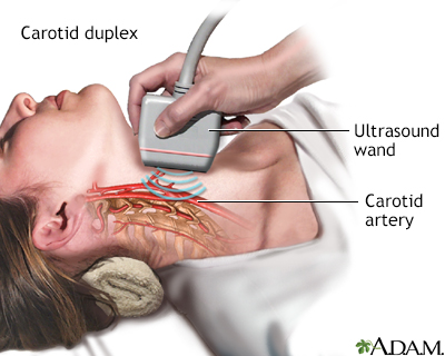
Carotid duplex is an ultrasound test that shows how well blood is flowing through the carotid arteries. The carotid arteries are located in the neck. They supply blood directly to the brain.
Images
How the Test is Performed
Ultrasound is a painless method that uses sound waves to create images of the inside of the body. The test is done in a vascular lab or radiology department.
The test is done in the following way:
- You lie on your back. Your head is supported to keep it from moving. The ultrasound technician applies a water-based gel to your neck to help with the transmission of the sound waves.
- Next, the technician moves a wand called a transducer back and forth over the area.
- The device sends sound waves to the arteries in your neck. The sound waves bounce off the blood vessels and form images or pictures of the insides of the arteries.
How to Prepare for the Test
No preparation is necessary.
How the Test will Feel
You may feel some pressure as the transducer is moved around your neck. The pressure should not cause any pain. You may also hear a whooshing sound. This is normal.
Why the Test is Performed
This test checks blood flow in the carotid arteries. It can detect:
- Blood clotting (thrombosis)
- Narrowing in the arteries (stenosis)
- Other causes of blockage in the carotid arteries
Your health care provider may order this test if:
- You have had a stroke or transient ischemic attack (TIA)
- You need a follow-up test because your carotid artery was found to be narrowed in the past or you have had surgery on the artery
- Your provider hears an abnormal sound called a bruit over the carotid neck arteries. This may mean the artery is narrowed.
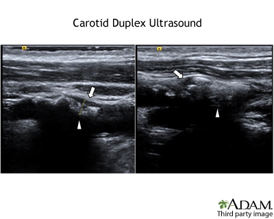
Normal Results
The results will tell your provider how open or narrowed your carotid arteries are. For example, the arteries may be 10% narrowed, 50% narrowed, or 75% narrowed.
A normal result means there is no problem with the blood flow in the carotid arteries. The artery is free of any significant blockage, narrowing, or other problem.
What Abnormal Results Mean
An abnormal result means the artery may be narrowed, or something is changing the blood flow in the carotid arteries. This is a sign of atherosclerosis or other blood vessel conditions.
In general, the more narrowed the artery is, the higher your risk for stroke.
Depending on the results, your provider may want you to:
- Consider treatment with medicines or surgery
- Have additional tests (such as cerebral angiography, CT angiography, and magnetic resonance angiography)
- Follow a healthy diet and lifestyle to prevent hardening of the arteries
- Repeat the test again in the future
Risks
There are no risks with having this procedure.
Related Information
UltrasoundBlood clots
Atherosclerosis
Carotid artery surgery - discharge
References
Adamczyx P, Liebeskind DS. Vascular imaging: computed tomographic angiography, magnetic resonance angiography, and ultrasound. In: Jankovic J, Mazziotta JC, Pomeroy SL, Newman NJ, eds. Bradley and Daroff's Neurology in Clinical Practice. 8th ed. Philadelphia, PA: Elsevier; 2022:chap 41.
Bluth EI, Johnson SI, Troxclair L. The extracranial cerebral vessels. In: Rumack CM, Levine D, eds. Diagnostic Ultrasound. 6th ed. Philadelphia, PA: Elsevier; 2024:chap 24.
Polak JF, Pellerito JS. Carotid sonography: protocol and technical considerations. In: Pellerito JS, Polak JF, eds. Introduction to Vascular Ultrasonography. 7th ed. Philadelphia, PA: Elsevier; 2020:chap 5.
BACK TO TOPReview Date: 8/19/2024
Reviewed By: Joseph V. Campellone, MD, Department of Neurology, Cooper Medical School at Rowan University, Camden, NJ. Review provided by VeriMed Healthcare Network. Also reviewed by David C. Dugdale, MD, Medical Director, Brenda Conaway, Editorial Director, and the A.D.A.M. Editorial team.

Health Content Provider
06/01/2025
|
A.D.A.M., Inc. is accredited by URAC, for Health Content Provider (www.urac.org). URAC's accreditation program is an independent audit to verify that A.D.A.M. follows rigorous standards of quality and accountability. A.D.A.M. is among the first to achieve this important distinction for online health information and services. Learn more about A.D.A.M.'s editorial policy, editorial process and privacy policy. A.D.A.M. is also a founding member of Hi-Ethics. This site complied with the HONcode standard for trustworthy health information from 1995 to 2022, after which HON (Health On the Net, a not-for-profit organization that promoted transparent and reliable health information online) was discontinued. |
The information provided herein should not be used during any medical emergency or for the diagnosis or treatment of any medical condition. A licensed medical professional should be consulted for diagnosis and treatment of any and all medical conditions. Links to other sites are provided for information only -- they do not constitute endorsements of those other sites. © 1997- 2025 A.D.A.M., a business unit of Ebix, Inc. Any duplication or distribution of the information contained herein is strictly prohibited.
 All rights reserved.