CSF oligoclonal banding
Cerebrospinal fluid - immunofixation
CSF oligoclonal banding is a test to look for inflammation-related proteins in the cerebrospinal fluid (CSF). CSF is the clear fluid that flows in the space around the spinal cord and brain.
Oligoclonal bands are proteins called immunoglobulins. The presence of these proteins indicates inflammation of the central nervous system. The presence of oligoclonal bands may point to a diagnosis of multiple sclerosis.
Images
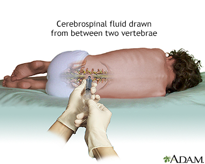
Presentation

How the Test is Performed
A sample of CSF is needed. A lumbar puncture (spinal tap) is the most common way to collect this sample.
Other methods for collecting CSF are rarely used, but may be recommended in some cases. They include:
- Cisternal puncture
- Ventricular puncture
- Removal of CSF from a tube that is already in the CSF, such as a shunt or ventricular drain.
After the sample is taken, it is sent to a lab for testing.
Why the Test is Performed
This test helps support the diagnosis of multiple sclerosis (MS). However, it does not confirm the diagnosis. Oligoclonal bands in the CSF may also be seen in other illnesses such as:
- Systemic lupus erythematosus
- Human immunodeficiency virus (HIV) infection
- Stroke
Normal Results
Normally, one or no bands should be found in the CSF.
Note: Normal value ranges may vary slightly among different laboratories. Talk to your health care provider about the meaning of your specific test results.
The examples above show the common measurements for results for these tests. Some laboratories use different measurements or may test different specimens.
What Abnormal Results Mean
There are two or more bandings found in the CSF and not in the blood. This may be a sign of multiple sclerosis or other diseases causing inflammation in the central nervous system.
Related Information
Multiple sclerosisReferences
Deluca GC, Griggs RC. Approach to the patient with neurologic disease. In: Goldman L, Schafer AI, eds. Goldman-Cecil Medicine. 26th ed. Philadelphia, PA: Elsevier; 2020:chap 368.
Karcher DS, McPherson RA. Cerebrospinal, synovial, serous body fluids, and alternative specimens. In: McPherson RA, Pincus MR, eds. Henry's Clinical Diagnosis and Management by Laboratory Methods. 24th ed. Philadelphia, PA: Elsevier; 2022:chap 30.
BACK TO TOPReview Date: 4/29/2023
Reviewed By: Joseph V. Campellone, MD, Department of Neurology, Cooper Medical School of Rowan University, Camden, NJ. Review provided by VeriMed Healthcare Network. Also reviewed by David C. Dugdale, MD, Medical Director, Brenda Conaway, Editorial Director, and the A.D.A.M. Editorial team.

Health Content Provider
06/01/2025
|
A.D.A.M., Inc. is accredited by URAC, for Health Content Provider (www.urac.org). URAC's accreditation program is an independent audit to verify that A.D.A.M. follows rigorous standards of quality and accountability. A.D.A.M. is among the first to achieve this important distinction for online health information and services. Learn more about A.D.A.M.'s editorial policy, editorial process and privacy policy. A.D.A.M. is also a founding member of Hi-Ethics. This site complied with the HONcode standard for trustworthy health information from 1995 to 2022, after which HON (Health On the Net, a not-for-profit organization that promoted transparent and reliable health information online) was discontinued. |
The information provided herein should not be used during any medical emergency or for the diagnosis or treatment of any medical condition. A licensed medical professional should be consulted for diagnosis and treatment of any and all medical conditions. Links to other sites are provided for information only -- they do not constitute endorsements of those other sites. © 1997- 2025 A.D.A.M., a business unit of Ebix, Inc. Any duplication or distribution of the information contained herein is strictly prohibited.
 All rights reserved.