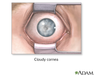
Cloudy cornea
Corneal opacification; Corneal scarring; Corneal edema
A cloudy cornea is a loss of transparency of the cornea.
Images
I Would Like to Learn About:
Causes
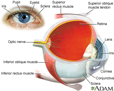
The cornea is the front wall of the eye. It is normally clear. It helps focus the light entering the eye.
Causes of cloudy cornea include:
- Inflammation of the cornea
- Sensitivity to non-infectious bacteria or toxins
- Infection of the cornea
- Keratitis
- Trachoma
- River blindness
- Corneal ulcers
- Swelling (edema)
- Acute glaucoma
- Birth injury
- Fuchs dystrophy
- Dryness of the eye due to Sjögren syndrome, vitamin A deficiency, or LASIK eye surgery
- Corneal dystrophy (inherited metabolic disease)
- Keratoconus
- Injury to the eye, including chemical burns and welding injury
- Tumors or growths on the eye
- Pterygium
- Bowen disease
Clouding may affect all or part of the cornea. It leads to different amounts of vision loss. You may not have any symptoms in the early stages.
Home Care
Contact your health care provider. There is no appropriate home care.
When to Contact a Medical Professional
Contact your provider if:
- The outer surface of your eye appears cloudy.
- You have trouble with your vision.
Note: You will need to see an ophthalmologist (eye doctor) for vision or eye problems. However, your primary care provider may also be involved if the problem could be due to a whole-body (systemic) disease.
What to Expect at Your Office Visit
The provider or eye doctor will examine your eyes and ask about your medical history. The two main questions will be if your vision is affected and if you have seen a spot on the front of your eye.
Other questions may include:
- When did you first notice this?
- Does it affect both eyes?
- Do you have trouble with your vision?
- Is it constant or intermittent?
- Do you wear contact lenses?
- Is there any history of injury to the eye?
- Has there been any discomfort? If so, is there anything that helps?
Tests may include:
- Biopsy of eyelid tissue
- Computer mapping of the cornea (corneal topography)
- Schirmer test for eye dryness
- Special photographs to measure the cells of the cornea
- Standard eye exam
- Ultrasound to measure corneal thickness
Related Information
IrisBlindness and vision loss
References
Cioffi GA, Liebmann JM. Diseases of the visual system. In: Goldman L, Cooney KA, eds. Goldman-Cecil Medicine. 27th ed. Philadelphia, PA: Elsevier; 2024:chap 391.
Kataguiri P, Kenyon KR. Corneal and external eye manifestations of systemic disease. In: Yanoff M, Duker JS, eds. Ophthalmology. 6th ed. Philadelphia, PA: Elsevier; 2023:chap 4.25.
Kuborn AM, Hassan SE. The impact of vision loss on attitudes toward autonomous vehicles: a vision-centric analysis. Optom Vis Sci. 2024 ;101(6):424-34. PMID: 38990241 pubmed.ncbi.nlm.nih.gov/38990241/.
Patel SS, Zaguia F, Goldstein DA. Episcleritis and scleritis. In: Yanoff M, Duker JS, eds. Ophthalmology. 6th ed. Philadelphia, PA: Elsevier; 2023:chap 4.11.
Wang EY, Kong X, Wolle M, et al. Global trends in blindness and vision impairment resulting from corneal opacity 1984–2020: A meta-analysis. Ophthalmology. 2023:130(8):863-71.PMID: 36963570 pubmed.ncbi.nlm.nih.gov/36963570/.
BACK TO TOPReview Date: 8/5/2024
Reviewed By: Franklin W. Lusby, MD, Ophthalmologist, Lusby Vision Institute, La Jolla, CA. Also reviewed by David C. Dugdale, MD, Medical Director, Brenda Conaway, Editorial Director, and the A.D.A.M. Editorial team.

Health Content Provider
06/01/2025
|
A.D.A.M., Inc. is accredited by URAC, for Health Content Provider (www.urac.org). URAC's accreditation program is an independent audit to verify that A.D.A.M. follows rigorous standards of quality and accountability. A.D.A.M. is among the first to achieve this important distinction for online health information and services. Learn more about A.D.A.M.'s editorial policy, editorial process and privacy policy. A.D.A.M. is also a founding member of Hi-Ethics. This site complied with the HONcode standard for trustworthy health information from 1995 to 2022, after which HON (Health On the Net, a not-for-profit organization that promoted transparent and reliable health information online) was discontinued. |
The information provided herein should not be used during any medical emergency or for the diagnosis or treatment of any medical condition. A licensed medical professional should be consulted for diagnosis and treatment of any and all medical conditions. Links to other sites are provided for information only -- they do not constitute endorsements of those other sites. © 1997- 2025 A.D.A.M., a business unit of Ebix, Inc. Any duplication or distribution of the information contained herein is strictly prohibited.
 All rights reserved.