Episcleritis
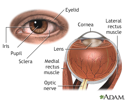
Episcleritis is irritation and inflammation of the episclera, a thin layer of tissue covering the white part (sclera) of the eye. It is not an infection.
Images
I Would Like to Learn About:
Causes
Episcleritis is a common condition. In most cases the problem is mild and vision is normal.
The cause is often unknown. But, it may occur with certain diseases, such as:
Symptoms
Symptoms include:
- A pink or purple color to the normally white part of the eye
- Eye pain
- Eye tenderness
- Sensitivity to light
- Tearing of the eye
Exams and Tests
Your health care provider or eye doctor will do an eye exam to diagnose the disorder. Most of the time, no special tests are needed.
Treatment
The condition most often goes away on its own in 1 to 2 weeks. Using corticosteroid eye drops may help ease the symptoms faster.
Outlook (Prognosis)
Episcleritis most often improves without treatment. However, treatment may make symptoms go away sooner.
Possible Complications
In some cases, the condition may return. Rarely, irritation and inflammation of the white part of the eye may develop. This is called scleritis.
When to Contact a Medical Professional
Contact your provider if you have symptoms of episcleritis that last for more than 2 weeks. Get checked again if your pain gets worse or you have problems with your vision.
Related Information
ScleraConjunctiva
Scleritis
Rheumatoid arthritis
Sjogren syndrome
Shingles
Pulmonary tuberculosis
References
Barry RJ, Denniston AK, Rhodes B, et al. Rheumatic disease. In: Sadda SVR, Sarraf D, Freund KB, et al, eds. Ryan's Retina. 7th ed. Philadelphia, PA: Elsevier; 2023:chap 81.
Cioffi GA, Liebmann JM. Diseases of the visual system. In: Goldman L, Cooney KA, eds. Goldman-Cecil Medicine. 27th ed. Philadelphia, PA: Elsevier; 2024:chap 391.
Patel SS, Zaguia F, Goldstein DA. Episcleritis and scleritis. In: Yanoff M, Duker JS, eds. Ophthalmology. 6th ed. Philadelphia, PA: Elsevier; 2023:chap 4.11.
Promelle V, Goeb V, Gueudry J. Rheumatoid arthritis associated episcleritis and scleritis: an update on treatment perspectives. J Clin Med. 2021;10(10):2118. PMID: 34068884 pubmed.ncbi.nlm.nih.gov/34068884/.
BACK TO TOPReview Date: 7/9/2024
Reviewed By: Audrey Tai, DO, MS, Athena Eye Care, Mission Viejo, CA. Also reviewed by David C. Dugdale, MD, Medical Director, Brenda Conaway, Editorial Director, and the A.D.A.M. Editorial team.

Health Content Provider
06/01/2025
|
A.D.A.M., Inc. is accredited by URAC, for Health Content Provider (www.urac.org). URAC's accreditation program is an independent audit to verify that A.D.A.M. follows rigorous standards of quality and accountability. A.D.A.M. is among the first to achieve this important distinction for online health information and services. Learn more about A.D.A.M.'s editorial policy, editorial process and privacy policy. A.D.A.M. is also a founding member of Hi-Ethics. This site complied with the HONcode standard for trustworthy health information from 1995 to 2022, after which HON (Health On the Net, a not-for-profit organization that promoted transparent and reliable health information online) was discontinued. |
The information provided herein should not be used during any medical emergency or for the diagnosis or treatment of any medical condition. A licensed medical professional should be consulted for diagnosis and treatment of any and all medical conditions. Links to other sites are provided for information only -- they do not constitute endorsements of those other sites. © 1997- 2025 A.D.A.M., a business unit of Ebix, Inc. Any duplication or distribution of the information contained herein is strictly prohibited.
 All rights reserved.