Peritonitis - secondary
Secondary peritonitis
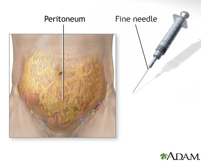
The peritoneum is the thin tissue that lines the inner wall of the abdomen and covers most of the organs in the abdomen. Peritonitis is present when this tissue becomes inflamed or infected. Secondary peritonitis is when another condition is the cause.
Images
I Would Like to Learn About:
Causes
Secondary peritonitis has several major causes.
- Bacteria may enter the peritoneum through a hole (perforation) in an organ of the digestive tract. The hole may be caused by a ruptured appendix, stomach ulcer, or perforated colon. It may also come from an injury, such as a gunshot or knife wound or following the ingestion of a sharp foreign body.
- Bile or chemicals released by the pancreas may leak into the abdominal cavity. This may be caused by swelling and inflammation of the pancreas called pancreatitis.
- Tubes or catheters placed into the abdomen may cause this problem. These include catheters for peritoneal dialysis, feeding tubes, and others.
An infection of the bloodstream (sepsis) may lead to an infection in the abdomen also. This is a severe illness.
This tissue may become infected when there is no clear cause.
Necrotizing enterocolitis occurs when the lining of the intestinal wall dies and can lead to peritonitis. This problem nearly always develops in an infant who is ill or born early.
Symptoms
Symptoms include:
- Swollen abdomen when your belly area is bigger than usual
- Abdominal pain
- Decreased appetite
- Fever
- Low urine output
- Nausea
- Thirst
- Vomiting
Note: There may be signs of shock.
Exams and Tests
During a physical exam, the health care provider may find abnormal vital signs with fever, rapid heart rate and breathing, low blood pressure, and a tender distended abdomen.
Tests may include:
- Blood culture
- Blood chemistry, including pancreatic enzymes
- Complete blood count
- Liver and kidney function tests
- X-rays or CT scan
- Peritoneal fluid culture, Gram stain and chemistry tests
- Urinalysis
Treatment
Often, surgery is needed to remove or treat sources of infection. These may be an infected bowel, an inflamed appendix, or an abscess or perforated diverticulum, usually due to diverticulitis.
General treatment includes:
- Antibiotics
- Fluids through a vein (IV)
- Pain medicines
- Tube through the nose into the stomach or intestine (nasogastric or NG tube)
Outlook (Prognosis)
The outcome can range from complete recovery to overwhelming infection and death. Factors that determine the outcome include:
- How long the symptoms were present before treatment began
- The person's general health
Possible Complications
Complications may include:
- Abscess
- Gangrene (dead) bowel requiring surgery
- Intraperitoneal adhesions (a potential cause of future bowel blockage)
- Septic shock
When to Contact a Medical Professional
Contact your provider if you have symptoms of peritonitis. This is a serious condition. It needs emergency treatment in most cases.
Related Information
BileBiliary system
Necrotizing enterocolitis
Septic shock
Adhesion
References
Mathews JB, Turaga K. Surgical peritonitis and other diseases of the peritoneum, mesentery, omentum, and diaphragm. In: Feldman M, Friedman LS, Brandt LJ, eds. Sleisenger and Fordtran's Gastrointestinal and Liver Disease. 11th ed. Philadelphia, PA: Elsevier; 2021:chap 39.
Privratsky AM, Barreto JC, Turnage RH, Mizell J, Badgwell B. Abdominal wall, umbilicus, peritoneum, mesenteries, omentum, and retroperitoneum. In: Townsend CM Jr, Beauchamp RD, Evers BM, Mattox KL, eds. Sabiston Textbook of Surgery. 21st ed. St Louis, MO: Elsevier; 2022:chap 44.
BACK TO TOPReview Date: 6/11/2024
Reviewed By: Jenifer K. Lehrer, MD, Department of Gastroenterology, Aria - Jefferson Health Torresdale, Jefferson Digestive Diseases Network, Philadelphia, PA. Review provided by VeriMed Healthcare Network. Also reviewed by David C. Dugdale, MD, Medical Director, Brenda Conaway, Editorial Director, and the A.D.A.M. Editorial team.

Health Content Provider
06/01/2025
|
A.D.A.M., Inc. is accredited by URAC, for Health Content Provider (www.urac.org). URAC's accreditation program is an independent audit to verify that A.D.A.M. follows rigorous standards of quality and accountability. A.D.A.M. is among the first to achieve this important distinction for online health information and services. Learn more about A.D.A.M.'s editorial policy, editorial process and privacy policy. A.D.A.M. is also a founding member of Hi-Ethics. This site complied with the HONcode standard for trustworthy health information from 1995 to 2022, after which HON (Health On the Net, a not-for-profit organization that promoted transparent and reliable health information online) was discontinued. |
The information provided herein should not be used during any medical emergency or for the diagnosis or treatment of any medical condition. A licensed medical professional should be consulted for diagnosis and treatment of any and all medical conditions. Links to other sites are provided for information only -- they do not constitute endorsements of those other sites. © 1997- 2025 A.D.A.M., a business unit of Ebix, Inc. Any duplication or distribution of the information contained herein is strictly prohibited.
 All rights reserved.