Cardiac intravascular ultrasound
IVUS; Ultrasound - coronary artery; Endovascular ultrasound; Intravascular echocardiography
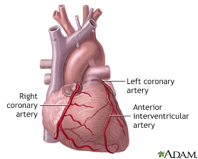
Intravascular ultrasound (IVUS) is a diagnostic test. This test uses sound waves to see inside blood vessels. It is useful for evaluating the coronary arteries that supply the heart.
Images


I Would Like to Learn About:
Description
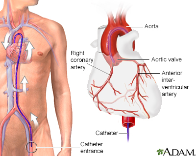
A tiny ultrasound wand is attached to the top of a thin tube. This tube is called a catheter. The catheter is inserted into an artery in your groin area and moved up to the heart. It is different from conventional duplex ultrasound. Duplex ultrasound is done from the outside of your body by placing the ultrasound transducer on the skin.
A computer measures how the sound waves reflect off blood vessels, and changes the sound waves into pictures. IVUS gives the health care provider a look at your coronary arteries from the inside-out.
IVUS is almost always done during a procedure. Reasons why it may be done include:
- Getting information about the heart or its blood vessels or to find out if you need heart surgery
- Treating some types of heart conditions
Angiography gives a general look at the coronary arteries. However, it can't show the walls of the arteries. IVUS images show the artery walls and can reveal cholesterol and fat deposits (plaques). Buildup of these deposits can increase your risk for a heart attack.
IVUS may help providers understand how stents become clogged. This is called stent restenosis.
Why the Procedure Is Performed
IVUS is commonly done to make sure a stent is correctly placed during angioplasty. It may also be done to determine where a stent should be placed.
IVUS may also be used to:
- View the aorta and structure of the artery walls, which can show plaque buildup
- Find which blood vessel is involved in aortic dissection
Risks
There is a slight risk for complications with angioplasty and cardiac catheterization. However, the tests are very safe when done by an experienced team. IVUS adds little additional risk.
Risks of anesthesia and surgery in general are:
- Reactions to medicines
- Breathing problems
- Bleeding, blood clots
- Infection
Other risks include:
- Damage to a heart valve or blood vessel
- Heart attack
- Irregular heartbeat (arrhythmia)
- Kidney failure (a higher risk in people who already have kidney problems or diabetes)
- Stroke (this is rare)
After the Procedure
After the test, the catheter is completely removed. A bandage is placed on the area. You will be asked to lie flat on your back with pressure on your groin area for a few hours after the test to prevent bleeding.
If IVUS was done during:
- Cardiac catheterization: You will stay in the hospital for about 3 to 6 hours.
- Angioplasty: You will stay in the hospital for 12 to 24 hours.
The IVUS does not add to the time you must stay in the hospital.
Related Information
UltrasoundCardiac catheterization
Stent
Coronary heart disease
References
Honda Y, Fitzgerald PJ, Yock PG. Intravascular ultrasound. In: Topol EJ, Teirstein PS, eds. Textbook of Interventional Cardiology. 8th ed. Philadelphia, PA: Elsevier; 2020:chap 65.
Yammine H, Ballast JK, Arko FR. Intravascular ultrasound. In: Sidawy AN, Perler BA, eds. Rutherford's Vascular Surgery and Endovascular Therapy. 10th ed. Philadelphia, PA: Elsevier; 2023:chap 32.
BACK TO TOPReview Date: 5/10/2024
Reviewed By: Neil Grossman, MD, Saint Vincent Radiological Associates, Framingham, MA. Review provided by VeriMed Healthcare Network. Also reviewed by David C. Dugdale, MD, Medical Director, Brenda Conaway, Editorial Director, and the A.D.A.M. Editorial team.

Health Content Provider
06/01/2025
|
A.D.A.M., Inc. is accredited by URAC, for Health Content Provider (www.urac.org). URAC's accreditation program is an independent audit to verify that A.D.A.M. follows rigorous standards of quality and accountability. A.D.A.M. is among the first to achieve this important distinction for online health information and services. Learn more about A.D.A.M.'s editorial policy, editorial process and privacy policy. A.D.A.M. is also a founding member of Hi-Ethics. This site complied with the HONcode standard for trustworthy health information from 1995 to 2022, after which HON (Health On the Net, a not-for-profit organization that promoted transparent and reliable health information online) was discontinued. |
The information provided herein should not be used during any medical emergency or for the diagnosis or treatment of any medical condition. A licensed medical professional should be consulted for diagnosis and treatment of any and all medical conditions. Links to other sites are provided for information only -- they do not constitute endorsements of those other sites. © 1997- 2025 A.D.A.M., a business unit of Ebix, Inc. Any duplication or distribution of the information contained herein is strictly prohibited.
