Fluorescein angiography
Retinal photography; Eye angiography; Angiography - fluorescein
Fluorescein angiography is an eye test that uses a special dye and camera to look at blood flow in the retina and choroid. These are the two layers in the back of the eye.
Images
I Would Like to Learn About:
How the Test is Performed
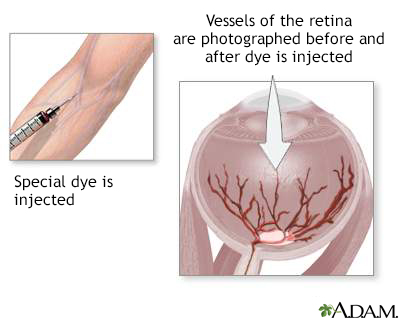
You will be given eye drops that make your pupil dilate. You will be asked to place your chin on a chin rest and your forehead against a support bar to keep your head still during the test.
The health care provider will take pictures of the inside of your eye including the back of your eye (retina). After the first group of pictures is taken, a dye called fluorescein is injected into a vein. Most often it is injected at the inside of your elbow. A camera-like device takes pictures as the dye moves through the blood vessels in the back of your eye.
A newer method called ultra-widefield fluorescein angiography can provide more information about certain diseases than regular fluorescein angiography.
How to Prepare for the Test
You will need someone to drive you home. Your vision may be blurry for up to 12 hours after the test.
You may be told to stop taking medicines that could affect the test results. Tell your provider about any allergies, particularly reactions to iodine.
You must sign an informed consent form. You must remove contact lenses before the test.
Tell the provider if you may be pregnant.
How the Test will Feel
When the needle is inserted, some people feel slight pain. Others feel only a prick or sting. Afterward, there may be some throbbing.
When the dye is injected, you may have mild nausea and a warm feeling in your body. These symptoms go away quickly most of the time.
The dye will cause your urine to be temporarily darker. It may be orange in color for a day or two after the test.
Why the Test is Performed
This test is done to see if there is proper blood flow in the blood vessels in the two layers in the back of your eye (the retina and choroid).
It can also be used to diagnose problems in the eye or to determine how well certain eye treatments are working.
Normal Results
A normal result means the vessels appear a normal size, there are no new abnormal vessels, and there are no blockages or leakages.
What Abnormal Results Mean
If blockage or leakage is present, the pictures will map the location for possible treatment.
An abnormal result of a fluorescein angiography may be due to:
- Blood flow (circulatory) problems, such as blockage of the arteries or veins
- Cancer
- Diabetic or other retinopathy
- High blood pressure
- Inflammation or swelling edema
- Macular degeneration
- Microaneurysms -- enlargement of capillaries in the retina
- Tumors
- Swelling of the optic disc
The test may also be done if you have:
Risks
There is a slight chance of infection any time the skin is broken. Rarely, a person is overly sensitive to the dye and may experience:
- Dizziness or faintness
- Dry mouth or increased salivation
- Hives
- Increased heart rate
- Metallic taste in mouth
- Nausea and vomiting
- Sneezing
Serious allergic reactions are rare.
Considerations
The test results are harder to interpret in people with cataracts. Blood flow problems shown on fluorescein angiography may suggest blood flow problems in other parts of the body.
Related Information
RetinaRetinopathy of prematurity
Diabetes and eye disease
High blood pressure and eye disease
Age-related macular degeneration
Retinal detachment
Retinal artery occlusion
Retinitis pigmentosa
References
Chen JJ, Peng M, Haug S, et al. Fluorescein angiography: basic principles and interpretation. In: Sadda SVR, Sarraf D, Freund KB, et al , eds. Ryan's Retina. 7th ed. Philadelphia, PA: Elsevier; 2023:chap 1.
Elnahry AG, Ramsey DJ. Automated image alignment for comparing microvascular changes detected by fluorescein angiography and optical coherence tomography angiography in diabetic retinopathy. Semin Ophthalmol. 2021;36(8):757-764. PMID: 33784213 pubmed.ncbi.nlm.nih.gov/33784213/.
Feinstein E, Olson JL, Mandava N. Camera-based ancillary retinal testing: autofluorescence, fluorescein, and indocyanine green angiography. In: Yanoff M, Duker JS, eds. Ophthalmology. 5th ed. Philadelphia, PA: Elsevier; 2019:chap 6.6.
Glacet-Bernard A, Miere A, Houmane B, Tilleul J, Souied E. Nonperfusion assessment in retinal vein occlusion: comparison between ultra-widefield fluorescein angiography and widefield optical coherence tomography angiography. Retina. 2021;41(6):1202-1209. PMID: 33105298 pubmed.ncbi.nlm.nih.gov/33105298/.
Karampelas M, Sim DA, Chu C, et al. Quantitative analysis of peripheral vasculitis, ischemia, and vascular leakage in uveitis using ultra-widefield fluorescein angiography. Am J Ophthalmol. 2015;159(6):1161-1168. PMID: 25709064 pubmed.ncbi.nlm.nih.gov/25709064/.
Taha NM, Asklany HT, Mahmoud AH, et al. Retinal fluorescein angiography: a sensitive and specific tool to predict coronary slow flow. Egypt Heart J. 2018;70(3):167-171. PMID: 30190642 pubmed.ncbi.nlm.nih.gov/30190642/.
BACK TO TOPReview Date: 8/22/2022
Reviewed By: Franklin W. Lusby, MD, Ophthalmologist, Lusby Vision Institute, La Jolla, CA. Also reviewed by David C. Dugdale, MD, Medical Director, Brenda Conaway, Editorial Director, and the A.D.A.M. Editorial team.

Health Content Provider
06/01/2025
|
A.D.A.M., Inc. is accredited by URAC, for Health Content Provider (www.urac.org). URAC's accreditation program is an independent audit to verify that A.D.A.M. follows rigorous standards of quality and accountability. A.D.A.M. is among the first to achieve this important distinction for online health information and services. Learn more about A.D.A.M.'s editorial policy, editorial process and privacy policy. A.D.A.M. is also a founding member of Hi-Ethics. This site complied with the HONcode standard for trustworthy health information from 1995 to 2022, after which HON (Health On the Net, a not-for-profit organization that promoted transparent and reliable health information online) was discontinued. |
The information provided herein should not be used during any medical emergency or for the diagnosis or treatment of any medical condition. A licensed medical professional should be consulted for diagnosis and treatment of any and all medical conditions. Links to other sites are provided for information only -- they do not constitute endorsements of those other sites. © 1997- 2024 A.D.A.M., a business unit of Ebix, Inc. Any duplication or distribution of the information contained herein is strictly prohibited.
 All rights reserved.