Ultrasound pregnancy
Pregnancy sonogram; Obstetric ultrasonography; Obstetric sonogram; Ultrasound - pregnancy; IUGR - ultrasound; Intrauterine growth - ultrasound; Polyhydramnios - ultrasound; Oligohydramnios - ultrasound; Placenta previa - ultrasound; Multiple pregnancy - ultrasound; Vaginal bleeding during pregnancy - ultrasound; Fetal monitoring - ultrasound
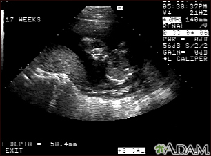
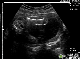
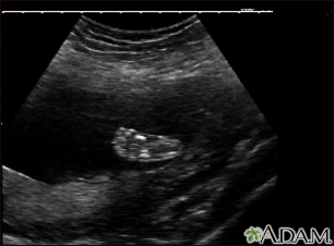
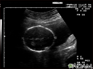
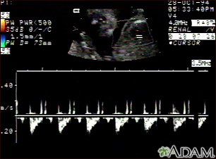
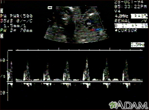
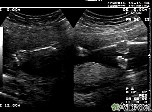
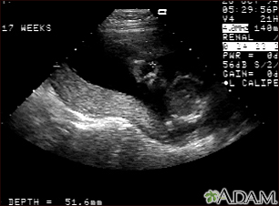
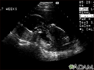
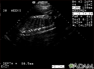
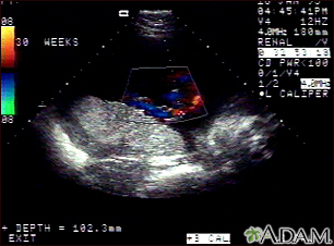
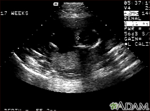
A pregnancy ultrasound is an imaging test that uses sound waves to create a picture of how a baby is developing in the womb (uterus). It is also used to check the female pelvic organs during pregnancy.
Images
Presentation

Animation

I Would Like to Learn About:
How the Test is Performed
To have the procedure:
- You will lie on your back on an exam table.
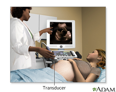
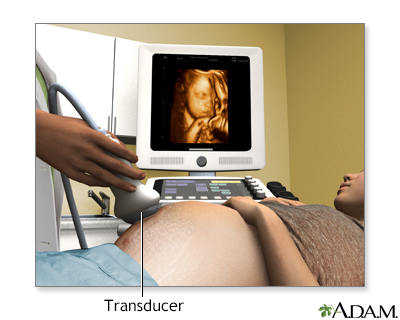
- The person performing the test will spread a clear, water-based gel on your belly and pelvis area. A handheld probe will then be moved over the area. The gel helps the probe transmit sound waves.
- These waves bounce off the body structures, including the developing baby, to create a picture on the ultrasound machine.
In some cases, a pregnancy ultrasound may be done by placing the probe into the vagina. This is more likely in early pregnancy. Many women will have the length of their cervix measured by vaginal ultrasonography around 20 to 24 weeks of pregnancy.
How to Prepare for the Test
You will need to have a full bladder to get the best ultrasound image. You may be asked to drink 2 to 3 glasses of liquid an hour before the test. DO NOT urinate before the procedure.
How the Test will Feel
There may be some discomfort from pressure on the full bladder. The conducting gel may feel slightly cold and wet. You will not feel the ultrasound waves.
Why the Test is Performed
An ultrasound may be done to determine if there is a problem with the pregnancy, how far along the pregnancy is, or to take measurements and screen for potential problems.
Talk to your health care provider to determine the most appropriate scanning schedule for you.
A pregnancy ultrasound may be done during the first 12 weeks of pregnancy to:
- Confirm a normal pregnancy
- Determine the baby's age
- Look for problems, such as ectopic pregnancies or the chances for a miscarriage
- Determine the baby's heart rate
- Look for multiple pregnancies (such as twins and triplets)
- Identify problems of the placenta, uterus, cervix, and ovaries
- Look for findings that might indicate an increased risk for Down syndrome
A pregnancy ultrasound may also be done in the second and third trimesters to:
- Determine the baby's age, growth, position, and sometimes sex.
- Identify any problems with how the fetus is developing.
- Look for twins or triplets. Look at the placenta, amniotic fluid, and pelvis.
Some centers are now performing a pregnancy ultrasound called a nuchal translucency screening test around 9 to 13 weeks of pregnancy. This test is done to look for signs of Down syndrome or other problems in the developing baby. This test is often combined with blood tests to improve the accuracy of results.
Ultrasound may also be performed to guide certain diagnostic procedures in the first and second trimesters to test the placenta and amniotic fluid for certain genetic disorders.
How many ultrasounds you will need depends on whether a previous scan or blood test has detected problems that require follow-up testing.
Normal Results
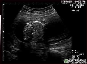
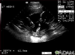
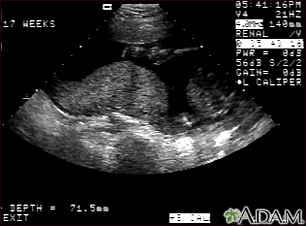
The developing baby, placenta, amniotic fluid, and surrounding structures appear normal for the gestational age.
Note: Normal results may vary slightly. Talk to your provider about the meaning of your specific test results.
What Abnormal Results Mean
Abnormal ultrasound results may be due to some of the following conditions:
- Birth defects
- Ectopic pregnancy
- Poor growth of a baby while in the mother's womb
- Multiple pregnancies
- Miscarriage
- Problems with the baby's position in the womb
- Problems with the placenta, including placenta previa and placental abruption
- Too little amniotic fluid
- Too much amniotic fluid (polyhydramnios)
- Tumors of pregnancy, including gestational trophoblastic disease
- Other problems with the ovaries, uterus, and remaining pelvic structures
Risks
Current ultrasound techniques appear to be safe. Ultrasound does not involve radiation.
Related Information
Ectopic pregnancyPolyhydramnios
Intrauterine growth restriction
References
Dugoff L, Wapner RJ. Prenatal diagnosis of congenital disorders. In: Lockwood CJ, Copel JA, Dugoff L, et al, eds. Creasy and Resnik's Maternal-Fetal Medicine: Principles and Practice. 9th ed. Philadelphia, PA: Elsevier; 2023:chap 30.
Richards DS. Obstetric ultrasound: imaging, dating, growth, and anomaly. In: Landon MB, Galan HL, Jauniaux ERM, et al, eds. Gabbe's Obstetrics: Normal and Problem Pregnancies. 8th ed. Philadelphia, PA: Elsevier; 2021:chap 9.
Wolf RB. Fetal abdominal imaging. In: Lockwood CJ, Copel JA, Dugoff L, et al, eds. Creasy and Resnik's Maternal-Fetal Medicine: Principles and Practice. 9th ed. Philadelphia, PA: Elsevier; 2023:chap 24.
BACK TO TOPReview Date: 3/31/2024
Reviewed By: LaQuita Martinez, MD, Department of Obstetrics and Gynecology, Emory Johns Creek Hospital, Alpharetta, GA. Also reviewed by David C. Dugdale, MD, Medical Director, Brenda Conaway, Editorial Director, and the A.D.A.M. Editorial team.

Health Content Provider
06/01/2025
|
A.D.A.M., Inc. is accredited by URAC, for Health Content Provider (www.urac.org). URAC's accreditation program is an independent audit to verify that A.D.A.M. follows rigorous standards of quality and accountability. A.D.A.M. is among the first to achieve this important distinction for online health information and services. Learn more about A.D.A.M.'s editorial policy, editorial process and privacy policy. A.D.A.M. is also a founding member of Hi-Ethics. This site complied with the HONcode standard for trustworthy health information from 1995 to 2022, after which HON (Health On the Net, a not-for-profit organization that promoted transparent and reliable health information online) was discontinued. |
The information provided herein should not be used during any medical emergency or for the diagnosis or treatment of any medical condition. A licensed medical professional should be consulted for diagnosis and treatment of any and all medical conditions. Links to other sites are provided for information only -- they do not constitute endorsements of those other sites. © 1997- 2025 A.D.A.M., a business unit of Ebix, Inc. Any duplication or distribution of the information contained herein is strictly prohibited.
 All rights reserved.