Ultrasound
Sonogram
Ultrasound uses high-frequency sound waves to make images of organs and structures inside the body.
Images
I Would Like to Learn About:
How the Test is Performed
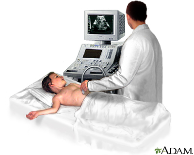
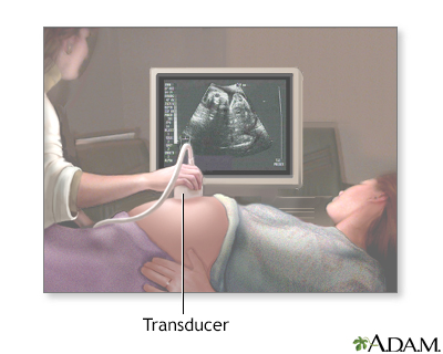
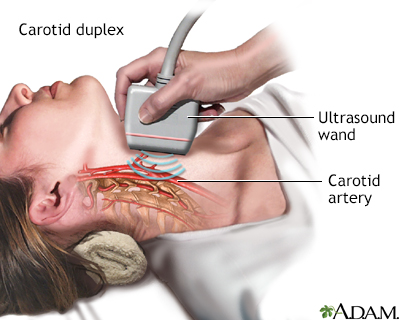
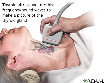
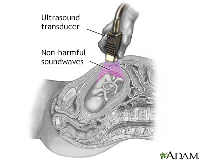
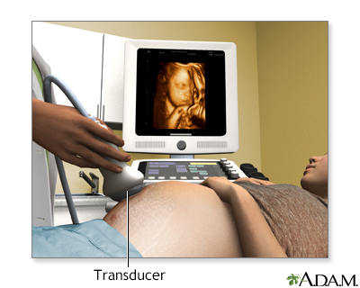
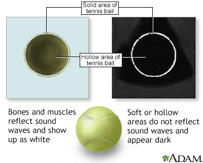
An ultrasound machine makes images so that organs inside the body can be examined. The machine sends out high-frequency sound waves, which reflect off body structures. A computer receives the waves and uses them to create a picture. Unlike with an x-ray or CT scan, this test does not use ionizing radiation.
The test is done in the ultrasound or radiology department.
- You will lie down for the test.
- A clear, water-based gel is applied to the skin on the area to be examined. The gel helps with the transmission of the sound waves.
- A handheld probe called a transducer is moved over the area being examined. You may need to change position so that other areas can be examined.
How to Prepare for the Test
Your preparation will depend on the part of the body being examined.
How the Test will Feel
Most of the time, ultrasound procedures do not cause discomfort. The conducting gel may feel a little cold and wet. You will feel the sonographer press the ultrasound probe against your body in the area they are reviewing.
Why the Test is Performed
The reason for the test will depend on your symptoms. An ultrasound test may be used to identify problems involving:
- Arteries in the neck
- Veins or arteries in the arms, legs, or abdomen
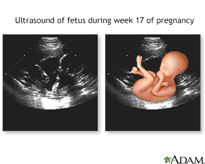
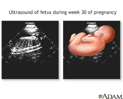
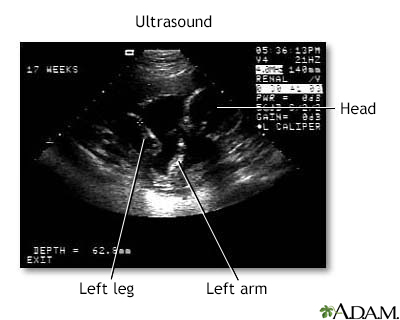
- Pregnancy
- Pelvis
- Abdomen and kidneys
- Breast
- Thyroid
- Eye and orbit
Normal Results
Results are considered normal if the organs and structures being examined look OK.
What Abnormal Results Mean
The meaning of abnormal results will depend on the part of the body being examined and the problem found. Talk to your health care provider about your questions and concerns.
Risks
There are no known risks. The test does not use ionizing radiation.
Considerations
Some types of ultrasound tests need to be done with a probe that is inserted into your body. Talk to your provider about how your test will be done.
Related Information
Cardiac intravascular ultrasoundReferences
Butts C. Ultrasound. In: Roberts JR, Custalow CB, Thomsen TW, eds. Roberts and Hedges' Clinical Procedures in Emergency Medicine and Acute Care. 7th ed. Philadelphia, PA: Elsevier; 2019:chap 66.
Fowler GC, Lefevre N. Emergency department, hospitalist, and office ultrasound (POCUS). In: Fowler GC, ed. Pfenninger and Fowler's Procedures for Primary Care. 4th ed. Philadelphia, PA: Elsevier; 2020:chap 214.
Morris AE, Adamson R, Frank J. Ultrasonography: principles and basic thoracic and vascular imaging. In: Broaddus VC, Ernst JD, King TE, et al, eds. Murray and Nadel's Textbook of Respiratory Medicine. 7th ed. Philadelphia, PA: Elsevier; 2022:chap 23.
Zhang D, Kahlili K, Yu H, Levine D. Physics of ultrasound. In: Rumack CM, Levine D, eds. Diagnostic Ultrasound. 6th ed. Philadelphia, PA: Elsevier; 2024:chap 1.
BACK TO TOPReview Date: 7/15/2024
Reviewed By: Jason Levy, MD, FSIR, Northside Radiology Associates, Atlanta, GA. Also reviewed by David C. Dugdale, MD, Medical Director, Brenda Conaway, Editorial Director, and the A.D.A.M. Editorial team.

Health Content Provider
06/01/2025
|
A.D.A.M., Inc. is accredited by URAC, for Health Content Provider (www.urac.org). URAC's accreditation program is an independent audit to verify that A.D.A.M. follows rigorous standards of quality and accountability. A.D.A.M. is among the first to achieve this important distinction for online health information and services. Learn more about A.D.A.M.'s editorial policy, editorial process and privacy policy. A.D.A.M. is also a founding member of Hi-Ethics. This site complied with the HONcode standard for trustworthy health information from 1995 to 2022, after which HON (Health On the Net, a not-for-profit organization that promoted transparent and reliable health information online) was discontinued. |
The information provided herein should not be used during any medical emergency or for the diagnosis or treatment of any medical condition. A licensed medical professional should be consulted for diagnosis and treatment of any and all medical conditions. Links to other sites are provided for information only -- they do not constitute endorsements of those other sites. © 1997- 2025 A.D.A.M., a business unit of Ebix, Inc. Any duplication or distribution of the information contained herein is strictly prohibited.
 All rights reserved.