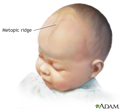
Metopic ridge
A metopic ridge is an abnormal shape of the skull. The ridge can be seen on the forehead.
Images


I Would Like to Learn About:
Considerations
The skull of an infant is made up of bony plates. The gaps between the plates allow for growth of the skull. The places where these plates connect are called sutures or suture lines. They do not fully close until the 2nd or 3rd year of life.
A metopic ridge occurs when the 2 bony plates in the front part of the skull join together too early.
The metopic suture remains unclosed throughout life in 1 in 10 people.
Causes
A birth defect called craniosynostosis is a common cause of metopic ridge. It can also be associated with other congenital skeletal defects.
When to Contact a Medical Professional
Contact your health care provider if you notice a ridge along your infant's forehead or a ridge forming on the skull.
The provider will perform a physical exam and ask questions about the child's medical history.
Tests may include:
- Head CT scan
- Skull x-ray
A metopic ridge must be differentiated from metopic synostosis, which is a more serious condition. Parents can find information and support at www.cappskids.org/metopic-ridge/.
No treatment or surgery is needed for a metopic ridge if it is the only skull abnormality.
Related Information
Sutures - ridgedReferences
Craniosynostosis and Positional Plagiocephaly Support (CAPPS) website. The metopic ridge / benign or surgical? www.cappskids.org/metopic-ridge/. Accessed January 28, 2022.
Gerety PA, Taylor JA, Bartlett SP. Nonsyndromic craniosynostosis. In: Rodriguez ED, Losee JE, Neligan PC, eds. Plastic Surgery: Volume 3: Craniofacial, Head and Neck Surgery and Pediatric Plastic Surgery. 4th ed. Philadelphia, PA: Elsevier; 2018:chap 32.
Jha RT, Magge SN, Keating RF. Diagnosis and surgical options for craniosynostosis. In: Ellenbogen RG, Sekhar LN, Kitchen ND, da Silva HB, eds. Principles of Neurological Surgery. 4th ed. Philadelphia, PA: Elsevier; 2018:chap 9.
Kinsman SL, Johnston MV. Congenital anomalies of the central nervous system. In: Kliegman RM, St. Geme JW, Blum NJ, Shah SS, Tasker RC, Wilson KM, eds. Nelson Textbook of Pediatrics. 21st ed. Philadelphia, PA: Elsevier; 2020:chap 609.
BACK TO TOPReview Date: 12/9/2021
Reviewed By: Franklin W. Lusby, MD, Ophthalmologist, Lusby Vision Institute, La Jolla, CA. Also reviewed by David Zieve, MD, MHA, Medical Director, Brenda Conaway, Editorial Director, and the A.D.A.M. Editorial team.

Health Content Provider
06/01/2025
|
A.D.A.M., Inc. is accredited by URAC, for Health Content Provider (www.urac.org). URAC's accreditation program is an independent audit to verify that A.D.A.M. follows rigorous standards of quality and accountability. A.D.A.M. is among the first to achieve this important distinction for online health information and services. Learn more about A.D.A.M.'s editorial policy, editorial process and privacy policy. A.D.A.M. is also a founding member of Hi-Ethics. This site complied with the HONcode standard for trustworthy health information from 1995 to 2022, after which HON (Health On the Net, a not-for-profit organization that promoted transparent and reliable health information online) was discontinued. |
The information provided herein should not be used during any medical emergency or for the diagnosis or treatment of any medical condition. A licensed medical professional should be consulted for diagnosis and treatment of any and all medical conditions. Links to other sites are provided for information only -- they do not constitute endorsements of those other sites. © 1997- 2024 A.D.A.M., a business unit of Ebix, Inc. Any duplication or distribution of the information contained herein is strictly prohibited.
