Pinguecula
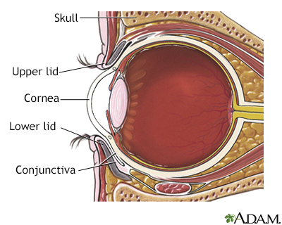
A pingueculum is a common, noncancerous growth of the conjunctiva. This is the clear, thin tissue that covers the white part of the eye (sclera). The growth occurs in the part of the conjunctiva that is exposed when the eye is open.
Images
I Would Like to Learn About:
Causes
The exact cause is unknown. Long-term sunlight exposure and eye irritation may be factors. Arc-welding is a major job-related risk.
Symptoms
A pingueculum looks like a small, yellowish bump on the conjunctiva near the cornea. It can appear on either side of the cornea. However, it more often occurs on the nose (nasal) side. The growth may increase in size, but usually over many years.
Exams and Tests
An eye exam is often enough to diagnose this disorder.
Treatment
The only treatment needed in most cases is the use of lubricating eye drops. Keeping the eye moist with artificial tears may help prevent the area from becoming inflamed. Temporary use of mild steroid eye drops can also be helpful. Rarely, the growth may need to be removed for comfort or for cosmetic reasons.
Outlook (Prognosis)
This condition is noncancerous (benign) and the outlook is good.
Possible Complications
The pingueculum may grow over the cornea and block vision. When this happens, the growth is called a pterygium. These two conditions occur under similar conditions. However, they are thought to be separate diseases.
When to Contact a Medical Professional
Contact your health care provider if the pingueculum changes in size, shape, or color, or if you would like to have it removed.
Prevention
Things you can do that may help prevent a pingueculum or keep the problem from getting worse include:
- Keeping the eye well lubricated with artificial tears
- Wearing good quality sunglasses
- Avoiding eye irritants
Related Information
MucosaConjunctiva
Eye burning - itching and discharge
References
American Academy of Ophthalmology website. Pinguecula and Pterygium. www.aao.org/eye-health/diseases/pinguecula-pterygium. Updated September 23, 2022. Accessed January 31, 2023.
Cioffi GA, Liebmann JM. Diseases of the visual system. In: Goldman L, Schafer AI, eds. Goldman-Cecil Medicine. 26th ed. Philadelphia, PA: Elsevier; 2020:chap 395.
Reidy JJ. Corneal and conjunctival degenerations. In: Mannis MJ, Holland EJ, eds. Cornea. 5th ed. Philadelphia, PA: Elsevier; 2022:chap 75.
Shtein RM, Sugar A. Pterygium and conjunctival degenerations. In: Yanoff M, Duker JS, eds. Ophthalmology. 6th ed. Philadelphia, PA: Elsevier; 2022:chap 4.9.
BACK TO TOPReview Date: 11/10/2022
Reviewed By: Franklin W. Lusby, MD, Ophthalmologist, Lusby Vision Institute, La Jolla, CA. Also reviewed by David C. Dugdale, MD, Medical Director, Brenda Conaway, Editorial Director, and the A.D.A.M. Editorial team.

Health Content Provider
06/01/2025
|
A.D.A.M., Inc. is accredited by URAC, for Health Content Provider (www.urac.org). URAC's accreditation program is an independent audit to verify that A.D.A.M. follows rigorous standards of quality and accountability. A.D.A.M. is among the first to achieve this important distinction for online health information and services. Learn more about A.D.A.M.'s editorial policy, editorial process and privacy policy. A.D.A.M. is also a founding member of Hi-Ethics. This site complied with the HONcode standard for trustworthy health information from 1995 to 2022, after which HON (Health On the Net, a not-for-profit organization that promoted transparent and reliable health information online) was discontinued. |
The information provided herein should not be used during any medical emergency or for the diagnosis or treatment of any medical condition. A licensed medical professional should be consulted for diagnosis and treatment of any and all medical conditions. Links to other sites are provided for information only -- they do not constitute endorsements of those other sites. © 1997- 2024 A.D.A.M., a business unit of Ebix, Inc. Any duplication or distribution of the information contained herein is strictly prohibited.
 All rights reserved.