A heart murmur is a blowing, whooshing, or rasping sound heard during a heartbeat. The sound is caused by turbulent (rough) blood flow through the heart valves or near the heart.
The valves of the heart open and close to control the flow of blood entering or leaving the heart.
Illustration
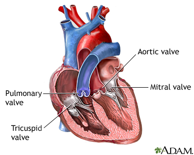
Heart valves - superior view
There are four valves located in the heart. Each valve either consists of two or three folds of thin tissue. When closed, the valve prevents blood from flowing backwards to its previous location. When open the valve allows blood to flow freely. Valve problems can occur because of congenital abnormalities, infection, or other causes.
Illustration
Heart valves - anterior view
There are four valves located in the heart. Each valve either consists of two or three folds of thin tissue. When closed, the valve prevents blood from flowing backwards to its previous location. When open the valve allows blood to flow freely. Valve problems can occur because of congenital abnormalities, infection, or other causes.
Illustration
Heart attack symptoms
Symptoms of a heart attack may widely vary, from the classic "elephant on the chest" feeling of crushing pain, to the nausea and heartburn mistaken for indigestion. In some patients, the symptoms may only be sudden fatigue or an oppressive feeling of impending doom.
Illustration
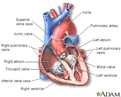
Heart chambers
The heart consists of four chambers in which blood flows. Blood enters the right atrium and passes through the right ventricle. The right ventricle pumps the blood to the lungs where it becomes oxygenated. The oxygenated blood is brought back to the heart by the pulmonary veins which enter the left atrium. From the left atrium blood flows into the left ventricle. The left ventricle pumps the blood to the aorta which will distribute the oxygenated blood to all parts of the body.
Illustration
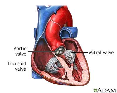
Heart - section through the middle
The interior of the heart is composed of valves, chambers, and associated vessels.
Illustration
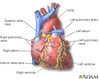
Heart - front view
The external structures of the heart include the ventricles, atria, arteries and veins. Arteries carry blood away from the heart while veins carry blood into the heart. The vessels colored blue indicate the transport of blood with relatively low content of oxygen and high content of carbon dioxide. The vessels colored red indicate the transport of blood with relatively high content of oxygen and low content of carbon dioxide.
Illustration
Heart bypass surgery incision
Minimally invasive heart bypass surgery is done without stopping the heart and putting the patient on a heart-lung machine. A 3 to 5 inch (8 to 13 cm) incision is made in the left part of the chest between the ribs. This incision is much less traumatic than the traditional heart bypass surgery incision which separates the breast bone. Minimally invasive heart bypass surgery allows the patient less pain and a faster recovery.
Illustration
Heart attack symptoms
Symptoms of a possible heart attack include chest pain and pain that radiates down the shoulder and arm. Some people (older adults, people with diabetes, and women) may have little or no chest pain. Or, they may experience unusual symptoms (shortness of breath, fatigue, weakness). Women are more likely than men to have symptoms of nausea, vomiting, back or jaw pain, and shortness of breath with chest pain.
Illustration
Heart beat
Two distinguishable sounds can be heard during the cycle of the beating heart when listened to with a stethoscope. The heart sounds are usually described as a lup-dup sound. These sounds are due to the closing of the valves of the heart. Unusual heart sounds are called murmurs.
Illustration
Heart transplant - series
Heart transplant - series
Presentation
Heart-lung transplant - series
Heart-lung transplant - series
Presentation
Heart valve surgery - series
Heart valve surgery - series
Presentation
Heart bypass surgery - series
Heart bypass surgery - series
Presentation
Heart bypass surgery - series
Heart bypass surgery - series
Presentation
Heartbeat - Animation
The heart has four chambers and four main blood vessels that either bring blood to the heart, or carry blood away. The four chambers are the right atrium and right ventricle and the left atrium and left ventricle. The blood vessels include the superior and inferior vena cava. These bring blood from the body to the right atrium. Next is the pulmonary artery that carries blood from the right ventricle to the lungs. The aorta is the body's largest artery. It carries oxygen-rich blood from the left ventricle to the rest of the body. Beneath the tough fibrous coating of the heart, you can see it beating. Inside the chambers are a series of one-way valves. These keep the blood flowing in one direction. Dye injected into the superior vena cava, will pass through all the heart's chambers during one cardiac cycle. Blood first enters the heart's right atrium. A muscle contraction forces the blood through the tricuspid valve into the right ventricle. When the right ventricle contracts, blood is forced through the pulmonary semilunar valve into the pulmonary artery. Then it travels to the lungs. In the lungs, the blood receives oxygen then leaves through the pulmonary veins. It returns to the heart and enters the left atrium. From there, blood is forced through the mitral valve into the left ventricle. This is the muscular pump that sends blood out to the rest of the body. When the left ventricle contracts, it forces blood through the aortic semilunar valve and into the aorta. The aorta and its branches carries the blood to all the body's tissues.
Heart formation - Animation
The embryo’s heart is the first organ that forms in its tiny body, and like most complex instruments, it begins with some simple structures. Let’s go back to 18 days after conception. . . Looking in the mother’s uterus, you can see the embryo surrounded by its yolk sac and amnion. Let’s take a look inside. Here’s a diagram of the embryo seen from a side view. Right now, it’s about the size of a raisin. There’s the head region and that red-colored area slightly above it contains two tubes that will form the embryo’s heart. Here’s what the tubes look like from a front view. On day 21, we see that the primitive heart tubes have moved below the embryo’s developing head region. And by day 22, the tubes have fused together, and have moved to the area that will eventually be our embryo’s thoracic, or chest cavity. It’s also about this time that the heart begins to beat for the first time. . . Here’s what it looks like from the front. Now let’s go back to day 18 and watch this happen from a different viewpoint. Here are two tubes in our embryo’s chest region seen from a front view. Watch this. . . Over the next two days, these tubes fuse together. Here’s another amazing part: the tube now starts bending and twisting and over the next 8 days it forms a simple version of the heart. By the time the embryo becomes a fetus at two months, the heart bears a close resemblance to what it will look like after the baby’s born. But the resemblance is only superficial. On the inside of the heart, things are much different in both form and function. Here’s a newborn heart on the left. Let’s take a closer look. There’s the right atrium right ventricle, left atrium and left ventricle. The two major blood vessels are the aorta and the pulmonary artery. The pathway of blood in the newborn heart works like this: oxygen-poor blood from the body enters the right atrium, then goes to the right ventricle. From the right ventricle, the blood is pumped to the lungs where it becomes oxygen rich. Then the blood flows back to the heart filling the left atrium and from there on to the left ventricle. The left ventricle pumps the oxygen rich blood through the aorta, which carries it to the rest of the newborn’s body. You can see the fetal heart has the same basic components as the newborn heart, but there are a couple important differences. Because the placenta is providing all of the oxygen the fetus requires, its lungs are not needed to perform this task, and therefore much of the fetus’ blood is detoured away from the lungs through two openings or connections. They are the foramen ovale, which connects the right and left atria, and the ductus arteriosus which connects the aorta and the pulmonary artery. As blood enters the heart into the right atrium some of the blood flows into the right ventricle as in the newborn, but also notice that some blood flows directly into the left atrium through the foramen ovale. This blood will pass directly into the left ventricle and be pumped out to the body without ever having gone to the lungs. In addition, some of the blood that did enter the right ventricle, and would normally go to the lungs, never reaches the lungs. Here lets watch. As blood is being pumped out of the right ventricle towards the lungs through the pulmonary artery, some of that blood escapes into the aorta through the ductus arteriosus, bypassing the lungs as it does. These two important connections will remain open up until the time of birth. Within thirty minutes after the baby’s first breath, the ductus arteriosus will completely close, and the flap of the foramen ovale will shut off like a valve. This happens because of an increase in pressure on the left side of the heart, and a decrease on the right side. These changes in the heart anatomy cause the blood to flow to the lungs, which will take over their lifelong job of supplying oxygen to the body. It’s incredible to think that this complex organ started off as a couple of tubes only 2 1/2 weeks ago.
Heartburn - Animation
Eating spicy foods, such as pizza, may cause a person to feel heartburn. Although the name may imply the heart, heartburn has nothing to do with the heart itself. Heartburn is pain felt in the chest by a burning sensation in the esophagus. Here, you can see the pizza passing from the mouth to the esophagus and on to the stomach. At the junction between the stomach and esophagus is the lower esophageal sphincter. This muscular sphincter acts as a valve that normally keeps food and stomach acid in the stomach, and prevents the stomach’s contents from regurgitating back into the esophagus. However, certain foods may affect the lower esophageal sphincter, making it less effective. That’s how heartburn begins. The stomach produces hydrochloric acid to digest food. The stomach has a mucous lining that protects it from hydrochloric acid, but the esophagus does not. So, when food and stomach acid regurgitate back into the esophagus, a burning feeling is felt near the heart. This feeling is known as heartburn. Antacids may be used to relieve heartburn by making stomach juices less acidic, thereby reducing the burning feeling felt in the esophagus. If heartburn becomes frequent or prolonged, medical intervention may be necessary to correct the problem.
Heart bypass surgery - Animation
Heart bypass surgery creates a new route, called a bypass, for blood and oxygen to reach the heart. Heart bypass surgery begins with an incision in the chest, and the breastbone is cut exposing the heart. Next, a portion of the saphenous vein, which is very large, is harvested from the inside of the leg. Pieces of this large vein are used to bypass the blocked coronary arteries, which are arteries that supply blood to the heart. The venous graft is sewn to the aorta, the main artery of the body, and to the affected coronary artery, to bypass the blocked site. The internal mammary artery from the chest may also be used to bypass a clogged artery. Several arteries may be bypassed depending on the condition of the heart. After the graft is created, the breastbone and chest are closed.
Heart attack - Animation
You feel a tight band of pain around your chest. The pain moves from your chest to your arms, shoulder, and neck. What could your pain mean? Could it be a heart attack. . . could it be the big one?Heart attacks are caused by interruption of blood supply to part of the heart. If the blood flow is blocked, your heart is starved of oxygen and heart cells die. A hard substance called plaque can build up in the walls of your coronary arteries. This plaque is made up of cholesterol and other cells. A heart attack can occur as a result of plaque buildup or the rupture of one of these plaques. We're not sure why heart attacks occur when they do. You may have a heart attack when you are resting or asleep, or after a sudden increase in physical activity, when you are outside in cold weather, or after a sudden, severe emotional or physical stress, including an illness. So, how is a heart attack treated?If you go to the hospital for a suspected heart attack, a doctor or nurse will listen to your chest with a stethoscope. You will have a blood test to look for heart damage. A coronary angiography test can show your doctor how well blood is moving through your heart. If blood moves slowly, or not at all through your coronary arteries, you have either a narrowed, or blocked artery. Other tests can look at the valves and chambers of your heart and check for abnormal heart rhythms. If you've had a heart attack, doctors can do an emergency procedure called angioplasty. This surgery or procedure can open narrowed or blocked blood vessels. Usually they'll place a small, metal mesh tube, called a stent, in your artery to help keep it open. You may also receive drugs to break up the clot in your artery. Sometimes, doctors will do heart bypass surgery to get blood flowing to your heart muscle again. After you are treated in the hospital for a heart attack, you may need to take medicines to thin your blood, to protect your heart, or to improve your cholesterol levels. You may need to take these medicines for the rest of your life. Most people who have had a heart attack also need cardiac rehabilitation. This will help you slowly increase your exercise level and learn how to follow a healthy lifestyle. After you have a heart attack, your chance of another is higher. How well you do after a heart attack depends on the damage to your heart and where the damage is, and what steps you take to prevent another one. If your heart can no longer pump blood to your body as well as it used to, you may have heart failure and will need lifelong treatment. Usually a person who has had a heart attack can slowly go back to normal activities, but you will need to take steps to prevent another heart attack.
Heart failure - Animation
If you cough a lot, often feel weak, have lost your appetite, and need to urinate a lot at night, you might have symptoms of heart failure. Heart failure is a long-term condition that usually comes on slowly. However, it can develop suddenly, for instance, after a heart attack. You have heart failure when your heart does not pump blood out of your heart very well, or when your heart muscles are stiff and do not easily fill up with blood. When you have heart failure, your heart cannot pump enough oxygen-rich blood to the rest of your body, especially when you exercise or move around a lot. As the heart loses the ability to pump blood, blood backs up in other parts of your body, including your lungs, liver, gastrointestinal tract, and your arms and legs. The most common cause of heart failure is coronary artery disease, the narrowing of the blood vessels that supply blood and oxygen to your heart. So, how do you know if you have heart failure?Get to your doctor. You may have trouble breathing, an irregular heartbeat, swollen legs, neck veins that stick out, and sounds from fluid built up in your lungs. Your doctor will check for these and other signs of heart failure. A test called an echocardiogram is often the best test to diagnose your heart failure. Your doctor can also use this test to find out why you have heart failure, and then monitor your condition going forward every three to six months. Your doctor will talk to you about knowing your body and symptoms that mean your heart failure is getting worse. You will need to learn to watch for changes in your heart rate, pulse, blood pressure, and weight. You will also need to limit salt in your diet, stop drinking alcohol, quit smoking if you need to, exercise, lose weight if you need to, and get enough rest. Your doctor will probably ask you to take medicines to treat your heart failure. These medicines can treat your symptoms, prevent your heart failure from getting worse, and help you live longer. If you have heart failure, taking your medicines, changing your lifestyle, and treating the condition that caused heart failure can go a long way toward improving your health. But heart failure is a chronic, or long-term, illness, which means it may get worse over time. Make sure you call your doctor if you start coughing more, have sudden weight gain or swelling, or feel week. Have someone take you to the emergency room right away if you have trouble with fainting, a fast and irregular heartbeat, or feel severe crushing chest pain.
What makes your heart beat? - Animation
Let’s take a closer look inside the heart. The yellow objects are not nerves. They’re actually specialized cardiac muscle cells in the walls of the heart. Their job is to send signals to the rest of the heart muscle and cause a contraction. Together, this group of cells is called the Cardiac conduction system. The main components of the Cardiac conduction system are the SA node, AV node, Bundle of His, Bundle branches, and Purkinje fibers. Let’s follow a signal through the contraction process. The SA node starts the sequence by causing the atrial muscles to contract. That’s why doctors sometimes call it the anatomical pacemaker. From there, the signal travels to the AV node, through the Bundle of His, down the Bundle branches, and through the Purkinje fibers, causing the ventricles to contract. This signal creates an electrical current that can be seen on a graph called an Electrocardiogram, or EKG. Doctors us an EKG as a way of seeing how well the Cardiac conduction system works. Any changes to the EKG can mean serious problems.
Review Date:
5/8/2024+
Reviewed By:
Thomas S. Metkus, MD, Assistant Professor of Medicine and Surgery, Johns Hopkins University School of Medicine, Baltimore, MD. Also reviewed by David C. Dugdale, MD, Medical Director, Brenda Conaway, Editorial Director, and the A.D.A.M. Editorial team.


, Heart valve disease with congestive heart failure, Severe congenital heart disease. Heart transplant surgery is not recommended for patients who have Kidney, lung, or liver disease, Insulin-dependent diabetes mellitus (IDDM), Other life-threatening diseases. </p>")
 transplant operations in the U.S. (over 1,500 cases per year). A healthy heart is obtained from a donor who has suffered brain death but remains on life-support. The healthy heart is transported in a special solution that preserves the organ. While the patient is deep asleep and pain-free (general anesthesia), an incision is made through the breast bone (sternum). The patient's blood is re-routed through tubes to a heart-lung bypass machine to keep the blood oxygen-rich and circulating. The patient's diseased heart is removed and the donor heart is stitched in place.</p>")


, an incision is made through the breast bone (sternum). Tubes are used to re-route the blood to a heart-lung bypass machine that keeps the blood oxygenated and circulating during the surgery.</p>")


 or leaking of the heart valve. Valve problems may be caused by infections (rheumatic fever) or birth defects and may cause heart failure (congestive heart failure) and infections (infective endocarditis). The surgery is done while the patient is in deep-sleep and pain-free (general anesthesia). An incision is made through the breast bone (sternum).</p>")

 or artificial (mechanical). Natural valves are from human donors (cadavers), modified natural valves are from animal donors (porcine, pigs) which are placed in synthetic rings, and artificial valves are made of metal or plastic. Natural valves rarely require life-long medication to prevent blood clot formation (anticoagulation), whereas artificial valves will require anticoagulation. The advantage of mechanical valves is that they last longer-thus, the tradeoff of lifelong anticoagulation in some cases is worth it to avoid a second valve replacement surgery.</p>")


 or heart bypass surgery is recommended when one or more coronary arteries are seriously blocked and blood supply to the heart muscle is insufficient. Several tests are done to identify the cause of the chest pain (angina), such as blood tests and x-ray studies (angiograms).</p>")

. It is done through an opening through the breast bone. While one surgeon is working on the chest, another surgeon works on taking a length of vein (saphenous vein) for the bypass through a long incision along the inside of the lower leg. The vein is sewn in above and below the blockage in the coronary artery. Alternatively, an artery from the interior aspect of the chest wall (internal mammary artery), or the arm (radial artery) is used.</p>")

. Chest tubes will be in place for the first 2 to 3 days to drain any residual blood and fluid from around the heart. Heart functions will be monitored. The full benefits from the operation may not be ascertained until 3 to 6 months after surgery. Sexual activity may be resumed 3 to 4 weeks after surgery. All activities that do not cause fatigue are permitted, but the patient must not strain the healing chest bone (sternum).</p>")
