X-ray
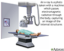
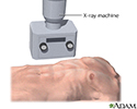
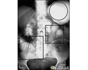
RadiographyX-rays are a type of electromagnetic radiation, just like visible light. An x-ray machine sends individual x-ray waves through the body. The images are recorded on a computer or film. Structures that are dense (such as bone) will block most of the x-ray, and will appear white. Metal and contrast media (special dye used to highlight areas of the...
X-ray
X-rays are a form of ionizing radiation that can penetrate the body to form an image on film. Structures that are dense (such as bone) will appear white, air will be black, and other structures will be shades of gray depending on density. X-rays can provide information about obstructions, tumors, and other diseases.
X-ray
illustration
X-ray
X-rays are a form of electromagnetic radiation, just like visible light. Structures that are dense (such as bone) will block most of the x-ray particles, and will appear white. Metal and contrast media (special dye used to highlight areas of the body) will also appear white. Structures containing air will be black and muscle, fat, and fluid will appear as shades of gray.
X-ray
illustration
Ileus - x-ray of bowel distension
This abdominal x-ray shows thickening of the bowel wall and swelling (distention) caused by a blockage (pseudo-obstruction) in the intestines. A solution containing a dye (barium), which is visible on X-ray, was swallowed by the patient (the procedure is known as an upper GI series).
Ileus - x-ray of bowel distension
illustration
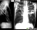
Tuberculosis, advanced - chest x-rays
Tuberculosis is an infectious disease that causes inflammation, the formation of tubercles and other growths within tissue, and can cause tissue death. These chest x-rays show advanced pulmonary tuberculosis. There are multiple light areas (opacities) of varying size that run together (coalesce). Arrows indicate the location of cavities within these light areas. The x-ray on the left clearly shows that the opacities are located in the upper area of the lungs toward the back. The appearance is typical for chronic pulmonary tuberculosis but may also occur with chronic pulmonary histiocytosis and chronic pulmonary coccidioidomycosis. Pulmonary tuberculosis is making a comeback with new resistant strains that are difficult to treat. Pulmonary tuberculosis is the most common form of the disease, but other organs can be infected.
Tuberculosis, advanced - chest x-rays
illustration
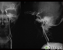
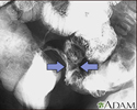
Carotid stenosis - X-ray of the left artery
A carotid arteriogram is an x-ray study designed to determine if there is narrowing or other abnormality in the carotid artery, a main artery to the brain. This is an angiogram of the left common carotid artery (both front-to-back and side views) showing a severe narrowing (stenosis) of the internal carotid artery just beyond the division of the common carotid artery into the internal and external branches.
Carotid stenosis - X-ray of the left artery
illustration
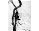
Carotid stenosis - X-ray of the right artery
This is an angiogram of the right carotid artery showing a severe narrowing (stenosis) of the internal carotid artery just past the carotid fork. There is enlargement of the artery or ulceration in the area after the stenosis in this close-up film. Note the narrowed segment toward the bottom of the picture.
Carotid stenosis - X-ray of the right artery
illustration
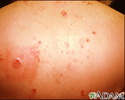
Multiple basal cell cancer due to x-ray therapy for acne
Basal cell carcinomas are more prevalent on sun or radiation exposed areas of skin. Here the typical lesion with raised, rolled, pearly borders with ulcerated center is seen on the back of a person previously irradiated for acne.
Multiple basal cell cancer due to x-ray therapy for acne
illustration
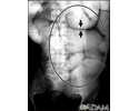
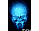
Eosinophilic granuloma - x-ray of the skull
This x-ray of the skull shows an eosinophilic granuloma (a lesion made-up of a type of white blood cell). This condition can range from a single eosinophilic granuloma to massive infiltration of skin, bone, and body organs.
Eosinophilic granuloma - x-ray of the skull
illustration
Ileus - x-ray of distended bowel and stomach
This abdominal X-ray shows a stomach filled with fluid and a swollen (distended) small bowel, caused by a blockage (pseudo-obstruction) in the intestines. A solution containing a dye (barium) that is visible on X-rays was swallowed by the patient (upper GI series).
Ileus - x-ray of distended bowel and stomach
illustration
Crohn disease - X-ray
This lower abdominal x-ray shows narrowing (stenosis) of the end of the small intestine (ileum), caused by Crohn disease. Crohn disease typically affects the small intestine, whereas ulcerative colitis typically affects the large intestine. A solution containing a dye (barium), was swallowed by the patient. When it passed into the small intestines, this x-ray was taken (lower GI series).
Crohn disease - X-ray
illustration
X-ray
X-rays are a form of ionizing radiation that can penetrate the body to form an image on film. Structures that are dense (such as bone) will appear white, air will be black, and other structures will be shades of gray depending on density. X-rays can provide information about obstructions, tumors, and other diseases.
X-ray
illustration
X-ray
X-rays are a form of electromagnetic radiation, just like visible light. Structures that are dense (such as bone) will block most of the x-ray particles, and will appear white. Metal and contrast media (special dye used to highlight areas of the body) will also appear white. Structures containing air will be black and muscle, fat, and fluid will appear as shades of gray.
X-ray
illustration
Ileus - x-ray of bowel distension
This abdominal x-ray shows thickening of the bowel wall and swelling (distention) caused by a blockage (pseudo-obstruction) in the intestines. A solution containing a dye (barium), which is visible on X-ray, was swallowed by the patient (the procedure is known as an upper GI series).
Ileus - x-ray of bowel distension
illustration
Tuberculosis, advanced - chest x-rays
Tuberculosis is an infectious disease that causes inflammation, the formation of tubercles and other growths within tissue, and can cause tissue death. These chest x-rays show advanced pulmonary tuberculosis. There are multiple light areas (opacities) of varying size that run together (coalesce). Arrows indicate the location of cavities within these light areas. The x-ray on the left clearly shows that the opacities are located in the upper area of the lungs toward the back. The appearance is typical for chronic pulmonary tuberculosis but may also occur with chronic pulmonary histiocytosis and chronic pulmonary coccidioidomycosis. Pulmonary tuberculosis is making a comeback with new resistant strains that are difficult to treat. Pulmonary tuberculosis is the most common form of the disease, but other organs can be infected.
Tuberculosis, advanced - chest x-rays
illustration
Carotid stenosis - X-ray of the left artery
A carotid arteriogram is an x-ray study designed to determine if there is narrowing or other abnormality in the carotid artery, a main artery to the brain. This is an angiogram of the left common carotid artery (both front-to-back and side views) showing a severe narrowing (stenosis) of the internal carotid artery just beyond the division of the common carotid artery into the internal and external branches.
Carotid stenosis - X-ray of the left artery
illustration
Carotid stenosis - X-ray of the right artery
This is an angiogram of the right carotid artery showing a severe narrowing (stenosis) of the internal carotid artery just past the carotid fork. There is enlargement of the artery or ulceration in the area after the stenosis in this close-up film. Note the narrowed segment toward the bottom of the picture.
Carotid stenosis - X-ray of the right artery
illustration
Multiple basal cell cancer due to x-ray therapy for acne
Basal cell carcinomas are more prevalent on sun or radiation exposed areas of skin. Here the typical lesion with raised, rolled, pearly borders with ulcerated center is seen on the back of a person previously irradiated for acne.
Multiple basal cell cancer due to x-ray therapy for acne
illustration
Eosinophilic granuloma - x-ray of the skull
This x-ray of the skull shows an eosinophilic granuloma (a lesion made-up of a type of white blood cell). This condition can range from a single eosinophilic granuloma to massive infiltration of skin, bone, and body organs.
Eosinophilic granuloma - x-ray of the skull
illustration
Ileus - x-ray of distended bowel and stomach
This abdominal X-ray shows a stomach filled with fluid and a swollen (distended) small bowel, caused by a blockage (pseudo-obstruction) in the intestines. A solution containing a dye (barium) that is visible on X-rays was swallowed by the patient (upper GI series).
Ileus - x-ray of distended bowel and stomach
illustration
Crohn disease - X-ray
This lower abdominal x-ray shows narrowing (stenosis) of the end of the small intestine (ileum), caused by Crohn disease. Crohn disease typically affects the small intestine, whereas ulcerative colitis typically affects the large intestine. A solution containing a dye (barium), was swallowed by the patient. When it passed into the small intestines, this x-ray was taken (lower GI series).
Crohn disease - X-ray
illustration
X-ray
RadiographyX-rays are a type of electromagnetic radiation, just like visible light. An x-ray machine sends individual x-ray waves through the body. The images are recorded on a computer or film. Structures that are dense (such as bone) will block most of the x-ray, and will appear white. Metal and contrast media (special dye used to highlight areas of the...
X-ray
RadiographyX-rays are a type of electromagnetic radiation, just like visible light. An x-ray machine sends individual x-ray waves through the body. The images are recorded on a computer or film. Structures that are dense (such as bone) will block most of the x-ray, and will appear white. Metal and contrast media (special dye used to highlight areas of the...
Review Date: 7/15/2024
Reviewed By: Jason Levy, MD, FSIR, Northside Radiology Associates, Atlanta, GA. Also reviewed by David C. Dugdale, MD, Medical Director, Brenda Conaway, Editorial Director, and the A.D.A.M. Editorial team.
 All rights reserved.