Health exams for: #AGEGROUP#
The following exams, tests, and procedures are recommended for #AGEGROUPLOWER#.#FEMALETEXT#
Select a link from the list below to learn how and why each test is performed, as well how to prepare for it.

The following exams, tests, and procedures are recommended for #AGEGROUPLOWER#.#FEMALETEXT#
Select a link from the list below to learn how and why each test is performed, as well how to prepare for it.
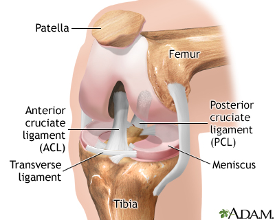
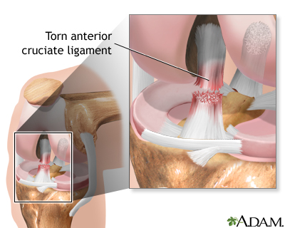
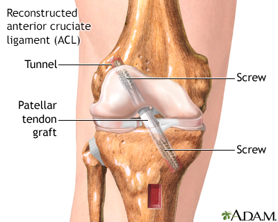
The knee is a complex joint made up of the distal end of the femur (femoral condyles) and the proximal end of the tibia (tibial plateau). A number of ligaments run between the femur and the tibia in the knee joint. The anterior cruciate ligament, the posterior cruciate ligament, and the meniscal ligaments are among the ligaments of the knee joint.
The knee is a complex joint made up of the distal end of the femur (femoral condyles) and the proximal end of the tibia (tibial plateau). A number of...
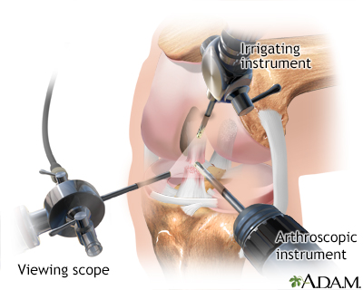
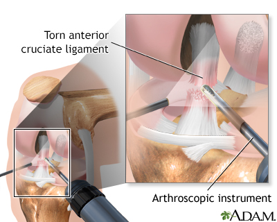
Injury to the ligaments of the knee are common sports-related injuries. Arthroscopy, which involves the use of a small camera and small instruments on the end of long narrow tubes, introduced into the knee through small incisions, may be recommended for knee problems such as A torn knee disc (meniscus); A damaged knee bone (patella); A damaged ligament; Inflamed or damaged lining of the joint (synovium).
Injury to the ligaments of the knee are common sports-related injuries. Arthroscopy, which involves the use of a small camera and small instruments ...
Several small punctures are made into the knee joint while the patient is deep asleep and pain-free (general anesthesia) or sleepy (sedated) and pain-free (regional anesthesia or spinal anesthesia).
Several small punctures are made into the knee joint while the patient is deep asleep and pain-free (general anesthesia) or sleepy (sedated) and pain...
The viewing scope (arthroscope) and other instruments are inserted into the knee joint. The surgeon can see the ligaments, the knee disc (meniscus), the knee bone (patella), the lining of the joint (synovium), and the rest of the joint. Damaged tissues can be removed. Arthroscopy can also be used to help view the inside of the knee while ligaments or tendons are repaired from the outside.
The viewing scope (arthroscope) and other instruments are inserted into the knee joint. The surgeon can see the ligaments, the knee disc (meniscus), ...
Patients are usually able to leave the hospital after arthroscopic knee surgery within 24 hours of surgery. The recovery time, and the need for physical therapy after surgery are determined by the injury treated and the procedure performed.
Patients are usually able to leave the hospital after arthroscopic knee surgery within 24 hours of surgery. The recovery time, and the need for physi...
Review Date: 4/24/2023
Reviewed By: C. Benjamin Ma, MD, Professor, Chief, Sports Medicine and Shoulder Service, UCSF Department of Orthopaedic Surgery, San Francisco, CA. Also reviewed by David C. Dugdale, MD, Medical Director, Brenda Conaway, Editorial Director, and the A.D.A.M. Editorial team.


