Health exams for: #AGEGROUP#
The following exams, tests, and procedures are recommended for #AGEGROUPLOWER#.#FEMALETEXT#
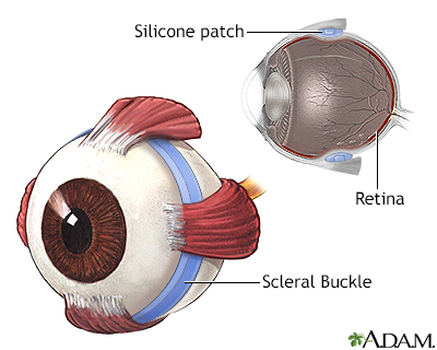
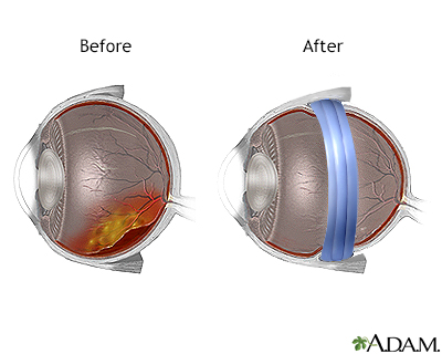
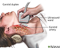
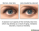
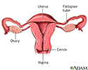
Select a link from the list below to learn how and why each test is performed, as well how to prepare for it.
Exam
Frequency

A.D.A.M. content is best viewed in IE9 or above, Firefox and Google Chrome browsers.
The information provided herein should not be used during any medical emergency or for the diagnosis or treatment of any medical condition. A licensed medical professional should be consulted for diagnosis and treatment of any and all medical conditions. Links to other sites are provided for information only -- they do not constitute endorsements of those other sites. No warranty of any kind, either expressed or implied, is made as to the accuracy, reliability, timeliness, or correctness of any translations made by a third-party service of the information provided herein into any other language. © 1997-
A.D.A.M., a business unit of Ebix, Inc. Any duplication or distribution of the information contained herein is strictly prohibited.
© 1997-
 All rights reserved.
All rights reserved.