Ultrasound, normal fetus - ventricles of brain
Ultrasound, normal fetus - ventricles of brain
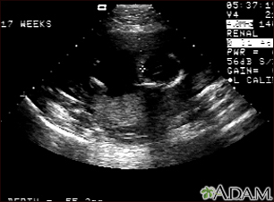
This is a normal fetal ultrasound performed at 17 weeks gestation. The development of the brain and nervous system begins early in fetal development. During an ultrasound, the technician usually looks for the presence of brain ventricles. Ventricles are spaces in the brain that are filled with fluid. In this early ultrasound, the ventricles can be seen as light lines extending through the skull, seen in the upper right side of the image. The cross hair is pointing to the front of the skull, and directly to the right, the lines of the ventricles are visible.
Review Date:
11/6/2023
Reviewed By
Neil K. Kaneshiro, MD, MHA, Clinical Professor of Pediatrics, University of Washington School of Medicine, Seattle, WA. Also reviewed by David C. Dugdale, MD, Medical Director, Brenda Conaway, Editorial Director, and the A.D.A.M. Editorial team.
Disclaimer
The information provided herein should not be used during any medical emergency or for the diagnosis or treatment of any medical condition. A licensed medical professional should be consulted for diagnosis and treatment of any and all medical conditions. Links to other sites are provided for information only -- they do not constitute endorsements of those other sites.
No warranty of any kind, either expressed or implied, is made as to the accuracy, reliability, timeliness, or correctness of any translations made by a third-party service of the information provided herein into any other language. © 1997-
A.D.A.M., a business unit of Ebix, Inc. Any duplication or distribution of the information contained herein is strictly prohibited.
 All rights reserved.