
Knee MRI scan
MRI - knee; Magnetic resonance imaging - kneeA knee MRI (magnetic resonance imaging) scan uses energy from strong magnets to create pictures of the knee joint and muscles and tissues.
An MRI does not use radiation (x-rays). Single MRI images are called slices. The images can be stored on a computer or printed on film. One exam produces many images.
How the Test is Performed
You will wear a hospital gown or clothes without metal zippers or snaps (such as sweatpants and a t-shirt). Please remove your watches, glasses, jewelry, and wallet. MRI can pull on any metal objects. Certain types of metal can cause blurry images.
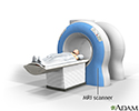
You will lie on a narrow table that slides into a large tunnel-like scanner.
Some exams use a special dye (contrast). Most of the time, you will get the dye through a vein (IV) in your arm or hand before the test. Sometimes, the dye is injected into a joint. The dye helps the radiologist see certain areas more clearly.
During the MRI, the person who operates the machine will watch you from another room. The test most often lasts 30 to 60 minutes, but may take longer. It can be loud. The technician can give you some ear plugs if needed.
How to Prepare for the Test
You may be asked not to eat or drink anything for 4 to 6 hours before the scan.
Tell your health care provider if you are afraid of closed spaces (have claustrophobia). You may be given a medicine to help you feel sleepy and less anxious. Your provider may suggest an "open" MRI, in which the machine is not as close to the body.
Before the test, tell your provider if you have:
- Brain aneurysm clips
- Certain types of artificial heart valves
- Heart defibrillator or pacemaker
- Inner ear (cochlear) implants
- Kidney disease or dialysis (you may not be able to receive contrast)
- Recently placed artificial joints or surgical repairs using metal plates and screws
- Certain types of vascular stents
Stents
A stent is a tiny tube placed into a hollow structure in your body. This structure can be an artery, a vein, or another structure, such as the tube ...
 ImageRead Article Now Book Mark Article
 - Worked with sheet metal in the past (you may need tests to check for metal pieces in your eyes)
Because the MRI contains strong magnets, metal objects are not allowed into the room with the MRI scanner:
- Pens, pocketknives, and eyeglasses may fly across the room.
- Items such as jewelry, watches, credit cards, and hearing aids can be damaged.
- Pins, hairpins, metal zippers, and similar metallic items can distort the images.
- Removable dental work should be taken out just before the scan.
How the Test will Feel
An MRI exam causes no pain. You will need to lie still. Too much movement can blur MRI images and cause errors.
The table may be hard or cold, but you can ask for a blanket or pillow. The machine makes loud thumping and humming noises when turned on. You can wear ear plugs to help block out the noise.
An intercom in the room allows you to speak to someone at any time. Some MRIs have televisions and special headphones to help the time pass.
There is no recovery time, unless you were given a medicine to relax. After an MRI scan, you can return to your normal diet, activity, and medicines.
Why the Test is Performed
Your provider may order this test if you have:
- An abnormal result on a knee x-ray or bone scan
- A feeling that your knee is giving away in the knee joint
- Buildup of joint fluid behind the knee (Baker cyst)
Baker cyst
Baker cyst is a buildup of joint fluid (synovial fluid) that forms a swelling behind the knee.
 ImageRead Article Now Book Mark Article
 - Fluid collecting in the knee joint
- Infection of the knee joint
- Knee cap injury
- Knee pain with fever
- Knee locking when you walk or moving
- Signs of damage to the knee muscle, cartilage, or ligaments
- Knee pain that does not get better with treatment
Knee pain
Knee pain is a common symptom in people of all ages. It may start suddenly, often after an injury or exercise. Knee pain also may begin as a mild d...
 ImageRead Article Now Book Mark Article
 - Instability of the knee
You may also have this test to check your progress after knee surgery.
Normal Results
A normal result means your knee looks OK.
What Abnormal Results Mean
Abnormal results may be due to a sprain or tear of the ligaments in the knee area.
Abnormal results may also be due to:
- Degeneration or changes that occur with age
- Meniscus or cartilage injuries
- Arthritis of the knee
- Avascular necrosis (also called osteonecrosis)
Avascular necrosis
Osteonecrosis is bone death caused by poor blood supply. It is most common in the hip and shoulder but can affect other large joints such as the kne...
 ImageRead Article Now Book Mark Article
 - Bone tumor or cancer
- Broken bone
- Buildup of joint fluid behind the knee (Baker cyst)
Baker cyst
Baker cyst is a buildup of joint fluid (synovial fluid) that forms a swelling behind the knee.
ImageRead Article Now Book Mark Article - Infection in the bone (osteomyelitis)
- Inflammation
- Injury of the knee cap
Talk to your provider if you have questions or concerns.
Risks
MRI uses no radiation. There have been no reported side effects from the magnetic fields and radio waves.
The most common type of contrast (dye) used is gadolinium. It is very safe. Allergic reactions to the substance are rare. However, gadolinium can be harmful to people with kidney problems that need dialysis. If you have kidney problems, please tell your provider before the test.
The strong magnetic fields created during an MRI can cause heart pacemakers and other implants to not work as well. It can also cause small pieces of metal inside your body to move or shift. For safety reasons, please do not bring anything that contains metal into the scanner room.
Considerations
Tests that may be done instead of a knee MRI include:
- CT scan of the knee
- Knee x-ray
Knee x-ray
This test is an x-ray of a knee, shoulder, hip, wrist, ankle, or other joint.
 ImageRead Article Now Book Mark Article
References
Helms CA. Magnetic resonance imaging of the knee. In: Helms CA, ed. Fundamentals of Skeletal Radiology. 5th ed. Philadelphia, PA: Elsevier; 2020:chap 9.
Kapoor G, Toms AP. Current status of imaging of the musculoskeletal system. In: Adam A, Dixon AK, Gillard JH, Schaefer-Prokop CM, eds. Grainger & Allison's Diagnostic Radiology. 7th ed. Philadelphia, PA: Elsevier; 2021:chap 38.
Riff AJ, Chalmers PN, Bach BR. Knee diagnosis and decision making. In: Miller MD, Thompson SR, eds. DeLee, Drez, & Miller's Orthopaedic Sports Medicine. 5th ed. Philadelphia, PA: Elsevier; 2020:chap 90.
MRI scans - illustration
MRI stands for magnetic resonance imaging. It allows imaging of the interior of the body without using x-rays or other types of ionizing radiation. An MRI scan is capable of showing fine detail of different tissues.
MRI scans
illustration
The structure of a joint - illustration
Joints, particularly hinge joints like the elbow and the knee, are complex structures made up of bone, muscles, synovium, cartilage, and ligaments that are designed to bear weight and move the body through space. The knee consists of the femur (thigh bone) above, and the tibia (shin bone) and fibula below. The kneecap (patella) glides through a shallow groove on the front part of the lower thigh bone. Ligaments and tendons connect the three bones of the knee, which are contained in the joint capsule (synovium) and are cushioned by cartilage.
The structure of a joint
illustration
Review Date: 4/24/2023
Reviewed By: C. Benjamin Ma, MD, Professor, Chief, Sports Medicine and Shoulder Service, UCSF Department of Orthopaedic Surgery, San Francisco, CA. Also reviewed by David C. Dugdale, MD, Medical Director, Brenda Conaway, Editorial Director, and the A.D.A.M. Editorial team.
 All rights reserved.