Magnetic resonance angiography
Magnetic resonance angiography (MRA) is an MRI exam of the blood vessels. Unlike traditional angiography that involves placing a tube (catheter) into the body, MRA is noninvasive.
MRI
A magnetic resonance imaging (MRI) scan is an imaging test that uses powerful magnets and radio waves to create pictures of the body. It does not us...

How the Test is Performed
You may be asked to wear a hospital gown. You can also wear clothing without metal fasteners (such as sweatpants and a t-shirt). Certain types of metal can cause blurry images.
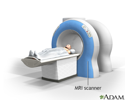
You will lie on a narrow table, which slides into a large tunnel-shaped scanner.
Some exams require a special dye (contrast). Most often, the dye is given before the test through a vein (IV) in your hand or forearm. The dye helps the radiologist see certain areas more clearly.
During the MRI, the person who operates the machine will watch you from another room. The test may take 1 hour or more.
How to Prepare for the Test
You may be asked not to eat or drink anything for 4 to 6 hours before the scan.
Tell your health care provider if you are afraid of close spaces (have claustrophobia). You may be given a medicine to help you feel sleepy and less anxious. Your provider may suggest an "open" MRI. In an open MRI, the machine is not as close to the body.
Before the test, tell your provider if you have:
- Brain aneurysm clips
- Artificial heart valve
- Heart defibrillator or pacemaker
Heart defibrillator
An implantable cardioverter-defibrillator (ICD) is a device that detects a life-threatening, rapid heartbeat. This abnormal heartbeat is called an a...
 ImageRead Article Now Book Mark Article
 - Inner ear (cochlear) implants
- Insulin pump or chemotherapy port
- Intrauterine device (IUD)
- Kidney disease or dialysis (you may not be able to receive contrast)
- Neurostimulator
- Recently placed artificial joints
- Vascular stent
Stent
A stent is a tiny tube placed into a hollow structure in your body. This structure can be an artery, a vein, or another structure, such as the tube ...
 ImageRead Article Now Book Mark Article
 - Worked with sheet metal in the past (you may need tests to check for tiny metal pieces in your eyes)
Because the MRI contains strong magnets, metal objects are not allowed into the room with the MRI scanner. Avoid carrying items such as:
- Pocketknives, pens, and eyeglasses
- Watches, credit cards, jewelry, and hearing aids
- Hairpins, metal zippers, pins, and similar items
- Removable dental implants
How the Test will Feel
An MRA exam causes no pain. If you have problems lying still or are very nervous, you may be given a medicine (sedative) to relax you. Moving too much can blur images and cause errors.
The table may be hard or cold, but you can ask for a blanket or pillow. The machine produces loud thumping and humming noises when turned on. You can wear ear plugs to help reduce the noise.
An intercom in the room allows you to speak to someone at any time. Some scanners have televisions and special headphones that you can use to help the time pass.
There is no recovery time, unless you were given a medicine to relax.
Why the Test is Performed
MRA is used to look at the blood vessels in all parts of the body. The test may be done of the head, heart, abdomen, lungs, kidneys, and legs.
It may be used to diagnose or evaluate conditions such as:
- Arterial aneurysm (an abnormal widening or ballooning of a part of an artery due to weakness in the wall of the blood vessel)
Arterial aneurysm
The aorta is the main blood vessel that supplies blood to the abdomen, pelvis, and legs. An abdominal aortic aneurysm (AAA) occurs when an area of t...
 ImageRead Article Now Book Mark Article
 - Aortic coarctation
Aortic coarctation
The aorta is a larger artery that carries blood from the heart to the vessels that supply the rest of the body with blood. If part of the aorta is n...
 ImageRead Article Now Book Mark Article
 - Aortic dissection
Aortic dissection
Aortic dissection is a serious condition in which there is a tear in the wall of the major artery carrying blood out of the heart (aorta). As the te...
ImageRead Article Now Book Mark Article - Stroke
Stroke
A stroke occurs when blood flow to a part of the brain stops. A stroke is sometimes called a "brain attack. " If blood flow is cut off for longer th...
 ImageRead Article Now Book Mark Article
 - Carotid artery disease
Carotid artery disease
Carotid artery disease causes the carotid arteries to become narrowed or blocked. The carotid arteries provide part of the main blood supply to your ...
 ImageRead Article Now Book Mark Article
 - Atherosclerosis of the arms or legs
Atherosclerosis
Atherosclerosis, sometimes called "hardening of the arteries," occurs when fat, cholesterol, and other substances build up in the walls of arteries. ...
 ImageRead Article Now Book Mark Article
 - Heart disease, including congenital heart disease
- Mesenteric artery ischemia
Mesenteric artery ischemia
Mesenteric artery ischemia occurs when there is a narrowing or blockage of one or more of the three major arteries that supply the small and large in...
 ImageRead Article Now Book Mark Article
 - Renal artery stenosis (narrowing of the blood vessels in the kidneys)
Normal Results
A normal result means the blood vessels do not show any signs of narrowing or blockage.
What Abnormal Results Mean
An abnormal result suggests a problem with one or more blood vessels. This may suggest:
- Atherosclerosis
Atherosclerosis
Atherosclerosis, sometimes called "hardening of the arteries," occurs when fat, cholesterol, and other substances build up in the walls of arteries. ...
ImageRead Article Now Book Mark Article - Trauma
- Congenital disease
- Other vascular condition
Risks
MRA is generally safe. It uses no radiation. To date, no side effects from the magnetic fields and radio waves have been reported.
The most common type of contrast used contains gadolinium. It is very safe. Allergic reactions to the substance rarely occur. However, gadolinium can be harmful to people with kidney problems who require dialysis. If you have kidney problems, please tell your provider before the test.
The strong magnetic fields created during an MRI can cause heart pacemakers and other implants to not work as well. They can also cause a piece of metal inside your body to move or shift.
Reviewed By
Neil Grossman, MD, Saint Vincent Radiological Associates, Framingham, MA. Review provided by VeriMed Healthcare Network. Also reviewed by David C. Dugdale, MD, Medical Director, Brenda Conaway, Editorial Director, and the A.D.A.M. Editorial team.
Carpenter JP, Litt H, Gowda M. Magnetic resonance imaging and arteriography. In: Sidawy AN, Perler BA, eds. Rutherford's Vascular Surgery and Endovascular Therapy. 10th ed. Philadelphia, PA: Elsevier; 2023:chap 30.
Kwong RY. Cardiovascular magnetic resonance imaging. In: Libby P, Bonow RO, Mann DL, Tomaselli GF, Bhatt DL, Solomon SD, eds. Braunwald's Heart Disease: A Textbook of Cardiovascular Medicine. 12th ed. Philadelphia, PA: Elsevier; 2022:chap 19.
 All rights reserved.