Virtual colonoscopy
Colonoscopy - virtual; CT colonography; Computed tomographic colonography; Colography - virtualA virtual colonoscopy (VC) is an imaging or x-ray test that looks for cancer, polyps, or other disease in the large intestine (colon). The medical name of this test is CT colonography.
Cancer
Cancer is the uncontrolled growth of abnormal cells in the body. Cancerous cells are also called malignant cells.
Read Article Now Book Mark ArticlePolyps
A colorectal polyp is a growth on the lining of the colon or rectum.

How the Test is Performed
A VC is different from a regular colonoscopy. A regular colonoscopy uses a long, lighted tool called a colonoscope that is inserted into the rectum and large intestine.
Colonoscopy
A colonoscopy is an exam that views the inside of the colon (large intestine) and rectum, using a tool called a colonoscope. The colonoscope has a sm...

A VC is done in the radiology department of a hospital or medical center. No sedatives are needed and no colonoscope is used.
The exam is done as follows:
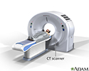
- You lie on your left side on a narrow table that is connected to a CT (computerized tomography) machine.
CT
A computed tomography (CT) scan is an imaging method that uses x-rays to create pictures of cross-sections of the body. Related tests include:Abdomin...
 ImageRead Article Now Book Mark Article
 - Your knees are drawn up toward your chest.
- A small, flexible tube is inserted into the rectum. Air is pumped through the tube to make the colon bigger and easier to see.
- You then lie on your back.
- The table slides into a large tunnel in the CT machine. X-rays of your colon are taken.
- X-rays are also taken while you lie on your stomach.
- You must stay very still during this procedure, because movement can blur the x-rays. You may be asked to hold your breath briefly while each x-ray is taken.
A computer combines all the images to form three-dimensional pictures of the colon. The radiologist can view the images on a video monitor.
How to Prepare for the Test
Your bowels need to be completely empty and clean for the exam. A problem in your large intestine that needs to be treated may be missed if your intestines are not cleaned out.
Your health care provider will give you the steps for cleansing your bowel. This is called bowel preparation. Steps may include:
- Using enemas
- Not eating solid foods for 1 to 3 days before the test
- Taking laxatives
You need to drink plenty of clear liquids for 1 to 3 days before the test. Examples of clear liquids are:
- Clear coffee or tea
- Fat-free bouillon or broth
- Gelatin
- Sports drinks
- Strained fruit juices
- Water
Keep taking your medicines unless your provider tells you otherwise.
You will need to stop taking iron pills or liquids a few days before the test, unless your provider tells you it is OK to continue. Iron can make your stool dark black. This makes it harder for the radiologist to view inside your bowel.
CT and MRI scanners are very sensitive to metals. Do not wear jewelry the day of your exam. You will be asked to change out of your street clothes and wear a hospital gown for the procedure.
How the Test will Feel
The x-rays are painless. Pumping air into the colon may cause cramping or gas pains.
After the exam:
- You may feel bloated and have mild abdominal cramping and pass a lot of gas.
- You should be able to return to your regular activities.
Why the Test is Performed
A VC may be done for the following reasons:
- Follow-up on colon cancer or polyps
Colon cancer
Colorectal cancer is cancer that starts in the large intestine (colon) or the rectum (end of the colon). It is also sometimes simply called colon ca...
 ImageRead Article Now Book Mark Article
 - Abdominal pain, changes in bowel movements, or weight loss
- Anemia due to low iron
Anemia
Anemia is a condition in which the body does not have enough healthy red blood cells. Red blood cells provide oxygen to body tissues. Different type...
 ImageRead Article Now Book Mark Article
 - Blood in the stool or black, tarry stools
- Screen for cancer of the colon or rectum (should be done every 5 years)
Your provider may recommend a regular colonoscopy instead of a VC. The reason is that a VC does not allow removing tissue samples or polyps.
Other times, a VC is done if a regular colonoscopy could not be completed.
Normal Results
Normal findings are images that show a healthy intestinal tract.
What Abnormal Results Mean
Abnormal test results may mean any of the following:
- Colorectal cancer
- Abnormal pouches on the lining of the intestines, called diverticulosis
Diverticulosis
Diverticula are small, bulging sacs or pouches that form on the inner wall of the intestine. Diverticulitis occurs when these pouches become inflame...
 ImageRead Article Now Book Mark Article
 - Colitis (a swollen and inflamed intestine) due to Crohn disease, ulcerative colitis, infection, or lack of blood flow
Colitis
Colitis is swelling (inflammation) of the large intestine (colon).
 ImageRead Article Now Book Mark Article
Crohn disease
Crohn disease is a disease where parts of the digestive tract become inflamed. It most often involves the lower end of the small intestine and the be...
 ImageRead Article Now Book Mark Article
Ulcerative colitis
Ulcerative colitis is a condition in which the lining of the large intestine (colon) and rectum become inflamed. It is a form of inflammatory bowel ...
 ImageRead Article Now Book Mark Article
 - Lower gastrointestinal (GI) bleeding
- Polyps
- Tumor
A regular colonoscopy may be done (on a different day) after a VC if:
- No cause for bleeding or other symptoms were found. A VC can miss some smaller problems in the colon.
- Problems that need a biopsy were seen on a VC.
Risks
Risks of a VC include:
- Exposure to radiation from the CT scan
- Nausea, vomiting, bloating, or rectal irritation from medicines used to prepare for the test
- Perforation of the intestine when the tube to pump air is inserted (extremely unlikely).
Considerations
The differences between a virtual and a conventional colonoscopy include:
- VC can view the colon from many different angles. This is not as easy with a regular colonoscopy.
- VC does not require sedation. You can usually go back to your normal activities right away after the test. A regular colonoscopy uses sedation and often the loss of a work day.
- VC using CT scanners expose you to radiation.
- Regular colonoscopy has a small risk for bowel perforation (creating a small tear). There is almost no such risk from a VC.
- VC is often not able to detect polyps smaller than 10 millimeters (mm). Regular colonoscopy can detect polyps of all sizes.
References
Garber JJ, Chung DC. Colonic polyps and polyposis syndromes. In: Feldman M, Friedman LS, Brandt LJ, eds. Sleisenger and Fordtran's Gastrointestinal and Liver Disease: Pathophysiology Diagnosis Management. 11th ed. Philadelphia, PA: Elsevier; 2021:chap 126.
Kim DH, Pickhardt PJ. Computed tomography colonography and evaluation of the colon. In: Gore RM, Levine MS, eds. Textbook of Gastrointestinal Radiology. 5th ed. Philadelphia, PA: Elsevier; 2021:chap 38.
National Cancer Institute website. Colorectal cancer prevention (PDQ) - health professional version. www.cancer.gov/types/colorectal/hp/colorectal-prevention-pdq. Updated August 18, 2023. Accessed July 26, 2024.
National Comprehensive Cancer Network website. NCCN clinical practice guidelines in oncology (NCCN guidelines): colorectal cancer screening. Version 1.2024 - February 27, 2024. www.nccn.org/professionals/physician_gls/pdf/colorectal_screening.pdf. Updated February 27, 2024. Accessed July 26, 2024.
Patel SG, May FP, Anderson JC, et al. Updates on age to start and stop colorectal cancer screening: recommendations from the U.S. Multi-Society Task Force on colorectal cancer. Gastroenterology. 2022;162(1):285-299. PMID: 34794816 pubmed.ncbi.nlm.nih.gov/34794816/.
Shaukat A, Kahi CJ, Burke CA, Rabeneck L, Sauer BG, Rex DK. ACG clinical guidelines: colorectal cancer screening 2021. Am J Gastroenterol. 2021;116(3):458-479. PMID: 33657038 pubmed.ncbi.nlm.nih.gov/33657038/.
US Preventive Services Task Force website. Final recommendation statement. Colorectal cancer: screening. www.uspreventiveservicestaskforce.org/uspstf/recommendation/colorectal-cancer-screening. Published May 18, 2021. Accessed February 11, 2024.
CT scan - illustration
CT stands for computerized tomography. In this procedure, a thin X-ray beam is rotated around the area of the body to be visualized. Using very complicated mathematical processes called algorithms, the computer is able to generate a 3-D image of a section through the body. CT scans are very detailed and provide excellent information for the physician.
CT scan
illustration
Review Date: 1/31/2023
Reviewed By: Michael M. Phillips, MD, Emeritus Professor of Medicine, The George Washington University School of Medicine, Washington, DC. Internal review and update on 07/26/2024 by David C. Dugdale, MD, Medical Director, Brenda Conaway, Editorial Director, and the A.D.A.M. Editorial team.


