
Chest x-ray
Chest radiography; Serial chest x-ray; X-ray - chestA chest x-ray is an x-ray of the chest, lungs, heart, large arteries, ribs, and diaphragm.
x-ray
X-rays are a type of electromagnetic radiation, just like visible light. An x-ray machine sends individual x-ray waves through the body. The images...

How the Test is Performed
You stand in front of the x-ray machine. You will be told to take a breath in and hold it when the x-ray is taken.
Two images are usually taken. You will first need to stand facing the machine, and then sideways.
How to Prepare for the Test
Tell your health care provider if you are pregnant. Chest x-rays are generally avoided during pregnancy, and special precautions are taken if they are needed.
How the Test will Feel
There is no discomfort. The imaging plate may feel cold.
Why the Test is Performed
Your provider may order a chest x-ray if you have any of the following symptoms:
- A persistent cough
-
Chest pain from a chest injury (with a possible rib fracture or lung complication) or from heart problems
Chest pain
Chest pain is discomfort or pain that you feel anywhere along the front of your body between your neck and upper abdomen.
 ImageRead Article Now Book Mark Article
 -
Coughing up blood
Coughing up blood
Coughing up blood is the spitting up of blood or bloody mucus from the lungs and throat (respiratory tract). Hemoptysis is the medical term for cough...
 ImageRead Article Now Book Mark Article
 -
Difficulty breathing
Difficulty breathing
Breathing difficulty may involve:Difficult breathing Uncomfortable breathingFeeling like you are not getting enough air
 ImageRead Article Now Book Mark Article
 - Fever
It may also be done if you have signs of tuberculosis, lung cancer, or other chest or lung diseases.
Tuberculosis
Pulmonary tuberculosis (TB) is a contagious bacterial infection that involves the lungs. It may spread to other organs.

Lung cancer
Lung cancer is cancer that starts in the lungs. The lungs are located in the chest. When you breathe, air goes through your nose, down your windpipe...
Lung diseases
Lung disease is any problem in the lungs that prevents the lungs from working properly. There are three main types of lung disease:Airway diseases -...

A serial chest x-ray is one that is repeated. It may be done to monitor changes found on a past chest x-ray.
What Abnormal Results Mean
Abnormal results may be due to many things, including:
In the lungs:
-
Collapsed lung
Collapsed lung
A collapsed lung occurs when air escapes from the lung. The air then fills the space outside of the lung between the lung and chest wall. This buil...
ImageRead Article Now Book Mark Article -
Collection of fluid around the lung
Collection of fluid around the lung
A pleural effusion is a buildup of fluid between the layers of tissue that line the lungs and chest cavity.
ImageRead Article Now Book Mark Article - Lung tumor (noncancerous or cancerous)
- Malformation of the blood vessels
-
Pneumonia
Pneumonia
Pneumonia is inflamed or swollen lung tissue due to infection with a germ. This article covers community-acquired pneumonia (CAP). This type of pneu...
 ImageRead Article Now Book Mark Article
 - Scarring of lung tissue
-
Tuberculosis
Tuberculosis
Pulmonary tuberculosis (TB) is a contagious bacterial infection that involves the lungs. It may spread to other organs.
ImageRead Article Now Book Mark Article -
Atelectasis
Atelectasis
Atelectasis is the collapse of part or, much less commonly, all of a lung.
 ImageRead Article Now Book Mark Article
In the heart:
- Problems with the size, position or shape of the heart
- Problems with the position, size and shape of the large arteries
- Evidence of heart failure
In the bones:
-
Fractures or other problems of the ribs and spine
Fractures
If more pressure is put on a bone than it can stand, it will split or break. A break of any size is called a fracture. If the broken bone punctures...
ImageRead Article Now Book Mark Article -
Osteoporosis
Osteoporosis
Osteoporosis is a disease in which bones become fragile and more likely to break (fracture).
 ImageRead Article Now Book Mark Article
In the mediastinum (middle part of the chest):
- Enlargement, which might be related to infection or tumor
Risks
There is low radiation exposure. X-rays are monitored and regulated to provide the minimum amount of radiation exposure needed to produce the image. Most experts feel that the benefits outweigh the risks. Pregnant women and children are more sensitive to the risks of x-rays.
References
Felker GM, Teerlink JR. Diagnosis and management of acute heart failure. In: Libby P, Bonow RO, Mann DL, Tomaselli GF, Bhatt DL, Solomon SD, eds. Braunwald's Heart Disease: A Textbook of Cardiovascular Medicine. 12th ed. Philadelphia, PA: Elsevier; 2022:chap 49.
Jokerst CE, Gotway MB. Thoracic radiology: noninvasive diagnostic imaging. In: Broaddus VC, Ernst JD, King TE, et al, eds. Murray and Nadel's Textbook of Respiratory Medicine. 7th ed. Philadelphia, PA: Elsevier; 2022:chap 20.
Nair A, Barnett JL, Semple TR. Current status of thoracic imaging. In: Adam A, Dixon AK, Gillard JH, Schaefer-Prokop CM, eds. Grainger & Allison's Diagnostic Radiology. 7th ed. Philadelphia, PA: Elsevier; 2021:chap 1.
-
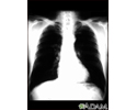
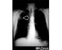
Aortic rupture - chest X-ray - illustration
Aortic rupture (a tear in the aorta, which is the major artery coming from the heart) can be seen on a chest X-ray. In this case, it was caused by a traumatic perforation of the thoracic aorta. This is how the X-ray appears when the chest is full of blood (right-sided hemothorax) seen here as cloudiness on the left side of the picture.
Aortic rupture - chest X-ray
illustration
-
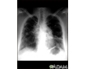
Lung cancer - frontal chest X-ray - illustration
A chest x-ray in a patient with central cancer of the right lung. Notice the white mass in the middle portion of the right lung (seen on the left side of the picture).
Lung cancer - frontal chest X-ray
illustration
-
Adenocarcinoma - chest x-ray - illustration
This chest x-ray shows adenocarcinoma of the lung. There is a rounded light spot in the right upper lung (left side of the picture) at the level of the second rib. The light spot has irregular and poorly defined borders and is not uniform in density. Diseases that may cause this type of x-ray result would be tuberculous or fungal granuloma, and malignant or benign tumors.
Adenocarcinoma - chest x-ray
illustration
-
Coal worker's lungs - chest x-ray - illustration
This chest x-ray shows coal worker's lungs. There are diffuse, small, light areas on both sides (1 to 3 mm) in all parts of the lungs. Diseases that may result in an x-ray like this include simple coal workers pneumoconiosis (CWP) - stage I, simple silicosis, miliary tuberculosis, histiocytosis X (eosinophilic granuloma), and other diffuse infiltrate pulmonary diseases.
Coal worker's lungs - chest x-ray
illustration
-
Coccidioidomycosis - chest X-ray - illustration
This chest x-ray shows the affects of a fungal infection, coccidioidomycosis. In the middle of the left lung (seen on the right side of the picture) there are multiple, thin-walled cavities (seen as light areas) with a diameter of 2 to 4 centimeters. To the side of these light areas are patchy light areas with irregular and poorly defined borders. Other diseases that may explain these x-ray findings include lung abscesses, chronic pulmonary tuberculosis, chronic pulmonary histoplasmosis, and others.
Coccidioidomycosis - chest X-ray
illustration
-
Coal workers pneumoconiosis - stage II - illustration
This chest x-ray shows stage II coal workers pneumoconiosis (CWP). There are diffuse, small light areas on both sides of the lungs. Other diseases that may explain these x-ray findings include simple silicosis, disseminated tuberculosis, metastatic lung cancer, and other diffuse, infiltrative pulmonary diseases.
Coal workers pneumoconiosis - stage II
illustration
-
Coal workers pneumoconiosis - stage II - illustration
This chest x-ray shows coal workers pneumoconiosis - stage II. There are diffuse, small (2 to 4 mm each), light areas throughout both lungs. In the right upper lung (seen on the left side of the picture), there is a light area (measuring approximately 2 cm by 4 cm) with poorly defined borders, representing coalescence (merging together) of previously distinct light areas. Diseases which may explain these x-ray findings include simple coal workers pneumoconiosis (CWP) - stage II, silico-tuberculosis, disseminated tuberculosis, metastatic lung cancer, and other diffuse infiltrative pulmonary diseases.
Coal workers pneumoconiosis - stage II
illustration
-
Coal workers pneumoconiosis, complicated - illustration
This picture shows complicated coal workers pneumoconiosis. There are diffuse, small, light areas (3 to 5 mm) in all areas on both sides of the lungs. There are large light areas which run together with poorly defined borders in the upper areas on both sides of the lungs. Diseases which may explain these X-ray findings include complicated coal workers pneumoconiosis (CWP), silico-tuberculosis, disseminated tuberculosis, metastatic lung cancer, and other diffuse infiltrative pulmonary diseases.
Coal workers pneumoconiosis, complicated
illustration
-
Coal workers pneumoconiosis, complicated - illustration
This picture shows complicated coal workers pneumoconiosis. There are diffuse, massive light areas that run together in the upper and middle parts of both lungs. These are superimposed on a background of small and poorly distinguishable light areas that are diffuse and located in both lungs. Diseases which may explain these x-ray findings include, but are not limited to complicated coal workers pneumoconiosis (CWP), silico-tuberculosis, and metastatic lung cancer.
Coal workers pneumoconiosis, complicated
illustration
-
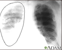
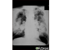
Tuberculosis, advanced - chest X-rays - illustration
Tuberculosis is an infectious disease that causes inflammation, the formation of tubercles and other growths within tissue, and can cause tissue death. These chest x-rays show advanced pulmonary tuberculosis. There are multiple light areas (opacities) of varying size that run together (coalesce). Arrows indicate the location of cavities within these light areas. The x-ray on the left clearly shows that the opacities are located in the upper area of the lungs toward the back. The appearance is typical for chronic pulmonary tuberculosis but may also occur with chronic pulmonary histiocytosis and chronic pulmonary coccidioidomycosis. Pulmonary tuberculosis is making a comeback with new resistant strains that are difficult to treat. Pulmonary tuberculosis is the most common form of the disease, but other organs can be infected.
Tuberculosis, advanced - chest X-rays
illustration
-
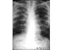
Pulmonary nodule - front view chest x-ray - illustration
This x-ray shows a single lesion (pulmonary nodule) in the upper right lung (seen as a light area on the left side of the picture). The nodule has distinct borders (well-defined) and is uniform in density. Tuberculosis (TB) and other diseases can cause this type of lesion.
Pulmonary nodule - front view chest x-ray
illustration
-
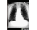
Sarcoid, stage II - chest X-ray - illustration
Sarcoid causes damage to the lung tissue that heals by scarring. The film shows a diffuse milky and granular appearance in the normally dark lung areas. This individual has marked decrease in lung function.
Sarcoid, stage II - chest X-ray
illustration
-
Sarcoid, stage IV - chest x-ray - illustration
This film shows advanced sarcoid, scarring of the lungs (the light streaking), and cavity formation (the dark areas in the upper right side of the picture).
Sarcoid, stage IV - chest x-ray
illustration
-
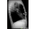
Pulmonary mass - side view chest X-ray - illustration
This individual has a mass in the upper part of the lung. Although the cause of the mass can be suspected, based on this person's history, there are many diseases that can produce lung lesions.
Pulmonary mass - side view chest X-ray
illustration
-
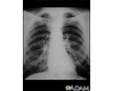
Bronchial cancer - chest X-ray - illustration
This is a chest x-ray of a person with bronchial cancer. This is a front view. The lungs are the two dark areas. The heart and other structures are white areas visible in the middle of the chest. The light areas that appear as subtle branches extending from the center into the lungs are cancerous.
Bronchial cancer - chest X-ray
illustration
-
Lung nodule, right middle lobe - chest X-ray - illustration
This is a chest X-ray (CXR) of a nodule in the right lung.
Lung nodule, right middle lobe - chest X-ray
illustration
-
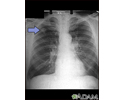
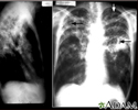
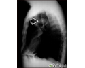
Lung mass, right upper lung - chest X-ray - illustration
This picture is a chest X-ray of a person with a lung mass. This is a front view, where the lungs are the two dark areas and the heart and other structures are visible in the middle of the chest. The X-ray shows a mass in the right upper lung, indicated with the arrow (seen on the left side of the picture).
Lung mass, right upper lung - chest X-ray
illustration
-
Lung nodule - front view chest X-ray - illustration
This is a chest X-ray showing mass in the right lower lung near heart (seen on the left side of the picture).
Lung nodule - front view chest X-ray
illustration
-
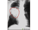
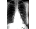
Aortic rupture - chest X-ray - illustration
Aortic rupture (a tear in the aorta, which is the major artery coming from the heart) can be seen on a chest X-ray. In this case, it was caused by a traumatic perforation of the thoracic aorta. This is how the X-ray appears when the chest is full of blood (right-sided hemothorax) seen here as cloudiness on the left side of the picture.
Aortic rupture - chest X-ray
illustration
-
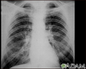
Lung cancer - frontal chest X-ray - illustration
A chest x-ray in a patient with central cancer of the right lung. Notice the white mass in the middle portion of the right lung (seen on the left side of the picture).
Lung cancer - frontal chest X-ray
illustration
-
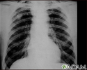
Adenocarcinoma - chest x-ray - illustration
This chest x-ray shows adenocarcinoma of the lung. There is a rounded light spot in the right upper lung (left side of the picture) at the level of the second rib. The light spot has irregular and poorly defined borders and is not uniform in density. Diseases that may cause this type of x-ray result would be tuberculous or fungal granuloma, and malignant or benign tumors.
Adenocarcinoma - chest x-ray
illustration
-
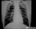
Coal worker's lungs - chest x-ray - illustration
This chest x-ray shows coal worker's lungs. There are diffuse, small, light areas on both sides (1 to 3 mm) in all parts of the lungs. Diseases that may result in an x-ray like this include simple coal workers pneumoconiosis (CWP) - stage I, simple silicosis, miliary tuberculosis, histiocytosis X (eosinophilic granuloma), and other diffuse infiltrate pulmonary diseases.
Coal worker's lungs - chest x-ray
illustration
-
Coccidioidomycosis - chest X-ray - illustration
This chest x-ray shows the affects of a fungal infection, coccidioidomycosis. In the middle of the left lung (seen on the right side of the picture) there are multiple, thin-walled cavities (seen as light areas) with a diameter of 2 to 4 centimeters. To the side of these light areas are patchy light areas with irregular and poorly defined borders. Other diseases that may explain these x-ray findings include lung abscesses, chronic pulmonary tuberculosis, chronic pulmonary histoplasmosis, and others.
Coccidioidomycosis - chest X-ray
illustration
-
Coal workers pneumoconiosis - stage II - illustration
This chest x-ray shows stage II coal workers pneumoconiosis (CWP). There are diffuse, small light areas on both sides of the lungs. Other diseases that may explain these x-ray findings include simple silicosis, disseminated tuberculosis, metastatic lung cancer, and other diffuse, infiltrative pulmonary diseases.
Coal workers pneumoconiosis - stage II
illustration
-
Coal workers pneumoconiosis - stage II - illustration
This chest x-ray shows coal workers pneumoconiosis - stage II. There are diffuse, small (2 to 4 mm each), light areas throughout both lungs. In the right upper lung (seen on the left side of the picture), there is a light area (measuring approximately 2 cm by 4 cm) with poorly defined borders, representing coalescence (merging together) of previously distinct light areas. Diseases which may explain these x-ray findings include simple coal workers pneumoconiosis (CWP) - stage II, silico-tuberculosis, disseminated tuberculosis, metastatic lung cancer, and other diffuse infiltrative pulmonary diseases.
Coal workers pneumoconiosis - stage II
illustration
-
Coal workers pneumoconiosis, complicated - illustration
This picture shows complicated coal workers pneumoconiosis. There are diffuse, small, light areas (3 to 5 mm) in all areas on both sides of the lungs. There are large light areas which run together with poorly defined borders in the upper areas on both sides of the lungs. Diseases which may explain these X-ray findings include complicated coal workers pneumoconiosis (CWP), silico-tuberculosis, disseminated tuberculosis, metastatic lung cancer, and other diffuse infiltrative pulmonary diseases.
Coal workers pneumoconiosis, complicated
illustration
-
Coal workers pneumoconiosis, complicated - illustration
This picture shows complicated coal workers pneumoconiosis. There are diffuse, massive light areas that run together in the upper and middle parts of both lungs. These are superimposed on a background of small and poorly distinguishable light areas that are diffuse and located in both lungs. Diseases which may explain these x-ray findings include, but are not limited to complicated coal workers pneumoconiosis (CWP), silico-tuberculosis, and metastatic lung cancer.
Coal workers pneumoconiosis, complicated
illustration
-
Tuberculosis, advanced - chest X-rays - illustration
Tuberculosis is an infectious disease that causes inflammation, the formation of tubercles and other growths within tissue, and can cause tissue death. These chest x-rays show advanced pulmonary tuberculosis. There are multiple light areas (opacities) of varying size that run together (coalesce). Arrows indicate the location of cavities within these light areas. The x-ray on the left clearly shows that the opacities are located in the upper area of the lungs toward the back. The appearance is typical for chronic pulmonary tuberculosis but may also occur with chronic pulmonary histiocytosis and chronic pulmonary coccidioidomycosis. Pulmonary tuberculosis is making a comeback with new resistant strains that are difficult to treat. Pulmonary tuberculosis is the most common form of the disease, but other organs can be infected.
Tuberculosis, advanced - chest X-rays
illustration
-
Pulmonary nodule - front view chest x-ray - illustration
This x-ray shows a single lesion (pulmonary nodule) in the upper right lung (seen as a light area on the left side of the picture). The nodule has distinct borders (well-defined) and is uniform in density. Tuberculosis (TB) and other diseases can cause this type of lesion.
Pulmonary nodule - front view chest x-ray
illustration
-
Sarcoid, stage II - chest X-ray - illustration
Sarcoid causes damage to the lung tissue that heals by scarring. The film shows a diffuse milky and granular appearance in the normally dark lung areas. This individual has marked decrease in lung function.
Sarcoid, stage II - chest X-ray
illustration
-
Sarcoid, stage IV - chest x-ray - illustration
This film shows advanced sarcoid, scarring of the lungs (the light streaking), and cavity formation (the dark areas in the upper right side of the picture).
Sarcoid, stage IV - chest x-ray
illustration
-
Pulmonary mass - side view chest X-ray - illustration
This individual has a mass in the upper part of the lung. Although the cause of the mass can be suspected, based on this person's history, there are many diseases that can produce lung lesions.
Pulmonary mass - side view chest X-ray
illustration
-
Bronchial cancer - chest X-ray - illustration
This is a chest x-ray of a person with bronchial cancer. This is a front view. The lungs are the two dark areas. The heart and other structures are white areas visible in the middle of the chest. The light areas that appear as subtle branches extending from the center into the lungs are cancerous.
Bronchial cancer - chest X-ray
illustration
-
Lung nodule, right middle lobe - chest X-ray - illustration
This is a chest X-ray (CXR) of a nodule in the right lung.
Lung nodule, right middle lobe - chest X-ray
illustration
-
Lung mass, right upper lung - chest X-ray - illustration
This picture is a chest X-ray of a person with a lung mass. This is a front view, where the lungs are the two dark areas and the heart and other structures are visible in the middle of the chest. The X-ray shows a mass in the right upper lung, indicated with the arrow (seen on the left side of the picture).
Lung mass, right upper lung - chest X-ray
illustration
-
Lung nodule - front view chest X-ray - illustration
This is a chest X-ray showing mass in the right lower lung near heart (seen on the left side of the picture).
Lung nodule - front view chest X-ray
illustration
Review Date: 8/19/2024
Reviewed By: Allen J. Blaivas, DO, Division of Pulmonary, Critical Care, and Sleep Medicine, VA New Jersey Health Care System, Clinical Assistant Professor, Rutgers New Jersey Medical School, East Orange, NJ. Review provided by VeriMed Healthcare Network. Also reviewed by David C. Dugdale, MD, Medical Director, Brenda Conaway, Editorial Director, and the A.D.A.M. Editorial team.
 All rights reserved.