
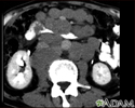
Abdominal CT scan
Computed tomography scan - abdomen; CT scan - abdomen; CT abdomen and pelvisAn abdominal CT scan is an imaging test that uses x-rays to create cross-sectional pictures of the belly area. CT stands for computed tomography.
How the Test is Performed
You will lie on a narrow table that slides into the center of the CT scanner. Most often, you will lie on your back with your arms raised above your head.
Once you are inside the scanner, the machine's x-ray beam rotates around you. Modern spiral scanners can perform the exam without stopping. There is very little noise.
x-ray
X-rays are a type of electromagnetic radiation, just like visible light. An x-ray machine sends individual x-ray waves through the body. The images...

A computer creates separate images of the belly area. These are called slices. These images can be stored, viewed on a monitor, printed on film or saved to a disk. Three-dimensional models of the belly area can be made by stacking the slices together.
You must be still during the exam, because movement causes blurred images. You may be told to hold your breath for short periods of time.
In many cases, an abdominal CT is done with a pelvis CT.
Pelvis CT
A computed tomography (CT) scan of the pelvis is an imaging method that uses x-rays to create cross-sectional pictures of the area between the hip bo...

The scan should take less than 30 minutes.
How to Prepare for the Test
You may need to have a special dye, called contrast, put into your body before some exams. Contrast helps certain areas show up better on the scans. Contrast can be administered in various ways. Such as:
- Contrast can be given through a vein (IV) in your hand or forearm. If contrast is used, you may also be asked not to eat or drink anything for 4 to 6 hours before the test.
- You may have to drink the contrast before the exam. When you drink it will depend on the type of exam being done. Contrast has a chalky taste, although some are flavored so they taste a little better. The contrast you drink will pass out of your body through your stools and is harmless.
- Rarely, the contrast may be given into your rectum using an enema.
Let your health care provider know if you have ever had a reaction to contrast. You may need to take medicines before the test to safely receive this substance.
Before receiving the contrast, tell your provider if you take the diabetes medicine metformin (Glucophage). People taking this medicine may have to stop taking it for a while before the test.
Let your provider know if you have any kidney problems. The IV contrast can worsen kidney function.
Too much weight can damage the scanner. Find out if the CT machine has a weight limit if you weigh more than 300 pounds (136 kg).
You will need to take off your jewelry and wear a hospital gown during the study.
How the Test will Feel
Lying on the hard table may be a bit uncomfortable.
If you have contrast through a vein (IV), you may have:
- Slight burning sensation
- Metallic taste in the mouth
- Warm flushing of the body
These feelings are normal and go away within a few seconds.
Why the Test is Performed
An abdominal CT scan makes detailed pictures of the structures inside your belly very quickly.
This test may be used to look for:
- Cause of blood in the urine
- Cause of abdominal pain or swelling
- Cause of abnormal blood test results such as liver or kidney problems
- Hernia
- Cause of a fever
- Masses and tumors, including cancer
- Infections or injury
- Kidney stones
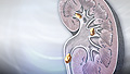
Kidney stones
A kidney stone is a solid mass made up of tiny crystals. One or more stones can be in the kidney or ureter at the same time.
 ImageRead Article Now Book Mark Article
 - Appendicitis
Appendicitis
Appendicitis is a condition in which your appendix gets inflamed. The appendix is a small pouch attached to the end of the large intestine.
 ImageRead Article Now Book Mark Article
What Abnormal Results Mean
The abdominal CT scan may show some cancers, including:
- Cancer of the renal pelvis or ureter
Cancer of the renal pelvis or ureter
Cancer of the renal pelvis or ureter is cancer that forms in the renal pelvis (center of the kidney) or ureter (tube that carries urine from the kidn...
 ImageRead Article Now Book Mark Article
 - Colon cancer
- Hepatocellular carcinoma
Hepatocellular carcinoma
Hepatocellular carcinoma is cancer that starts in the liver.
 ImageRead Article Now Book Mark Article
 - Lymphoma
- Melanoma
- Ovarian cancer
- Pancreatic cancer
Pancreatic cancer
Pancreatic cancer is cancer that starts in the pancreas.
ImageRead Article Now Book Mark Article - Pheochromocytoma
Pheochromocytoma
Pheochromocytoma is a rare tumor of adrenal gland tissue that typically arises from the adrenal gland. It results in the release of too much epineph...
 ImageRead Article Now Book Mark Article
 - Renal cell carcinoma (kidney cancer)
Renal cell carcinoma
Renal cell carcinoma is a type of kidney cancer that starts in the lining of very small tubes (tubules) in the kidney.
ImageRead Article Now Book Mark Article - Spread of cancers that began outside the belly
The abdominal CT scan may show problems with the gallbladder, liver, or pancreas, including:
- Acute cholecystitis
Acute cholecystitis
Acute cholecystitis is sudden swelling and irritation of the gallbladder. It causes severe belly pain.
ImageRead Article Now Book Mark Article - Alcoholic liver disease
- Cholelithiasis
Cholelithiasis
Gallstones are hard deposits that form inside the gallbladder. These may be as small as a grain of sand or as large as a golf ball.
ImageRead Article Now Book Mark Article - Pancreatic abscess
Pancreatic abscess
A pancreatic abscess is an area filled with pus within the pancreas.
ImageRead Article Now Book Mark Article - Pancreatic pseudocyst
Pancreatic pseudocyst
A pancreatic pseudocyst is a fluid-filled sac in the abdomen that arises from the pancreas. It may also contain tissue from the pancreas, enzymes, a...
ImageRead Article Now Book Mark Article - Pancreatitis
- Blockage of bile ducts
The abdominal CT scan may reveal the following kidney problems:
- Blockage of the kidneys
- Hydronephrosis (kidney swelling from the backflow of urine)
Hydronephrosis
Hydronephrosis is swelling of one kidney due to a backup of urine. This problem may occur in one kidney.
 ImageRead Article Now Book Mark Article
 - Kidney infection
- Kidney stones
- Kidney or ureter damage
- Polycystic kidney disease
Polycystic kidney disease
Polycystic kidney disease (PKD) is a kidney disorder passed down through families. In this disease, many cysts form in the kidneys, causing them to ...
 ImageRead Article Now Book Mark Article
Abnormal results may also be due to:
- Abdominal aortic aneurysm
Abdominal aortic aneurysm
The aorta is the main blood vessel that supplies blood to the abdomen, pelvis, and legs. An abdominal aortic aneurysm (AAA) occurs when an area of t...
 ImageRead Article Now Book Mark Article
 - Abscesses
Abscesses
An abscess is a collection of pus in any part of the body. In most cases, the area around an abscess is swollen and inflamed.
 ImageRead Article Now Book Mark Article
 - Appendicitis
Appendicitis
Appendicitis is a condition in which your appendix gets inflamed. The appendix is a small pouch attached to the end of the large intestine.
ImageRead Article Now Book Mark Article - Bowel wall thickening
Bowel wall thickening
Irritable bowel syndrome (IBS) is a disorder that leads to pain in abdomen and changes in bowel movements. IBS is not the same as inflammatory bowel...
ImageRead Article Now Book Mark Article - Crohn disease
- Renal artery stenosis
- Renal vein thrombosis
Renal vein thrombosis
Renal vein thrombosis is a blood clot that develops in the vein that drains blood from the kidney.
ImageRead Article Now Book Mark Article
Risks
Risks of CT scans include:
- Allergy to contrast dye
- Exposure to radiation
- Damage to kidney function from contrast dye
CT scans expose you to more radiation than regular x-rays. Many x-rays or CT scans over time may increase your risk for cancer. However, the risk from any one scan is small. Most modern scanners are able to reduce the radiation exposure. Talk to your provider about this risk and the benefit of the test for getting a correct diagnosis of your medical problem.
Some people have allergies to contrast dye. Let your provider know if you have ever had an allergic reaction to injected contrast dye.
The most common type of contrast given into a vein contains iodine. If you have an iodine allergy, you may have nausea or vomiting, sneezing, itching, or hives if you get this type of contrast. If you must be given such contrast, your provider may give you antihistamines (such as Benadryl) or steroids before the test.
Nausea or vomiting
Nausea is feeling an urge to vomit. It is often called "being sick to your stomach. "Vomiting or throwing-up forces the contents of the stomach up t...
Sneezing
A sneeze is a sudden, forceful, uncontrolled burst of air through the nose and mouth.

Itching
Itching is a tingling or irritation of the skin that makes you want to scratch the area. Itching may occur all over the body or only in one location...

Hives
Hives are raised, often itchy, red bumps (welts) on the surface of the skin. They can be an allergic reaction to food or medicine. They can also ap...

Antihistamines
An allergy is an immune response, or reaction, to substances (allergens) that are usually not harmful. In someone with allergies, the immune respons...

Your kidneys help remove IV dye from the body. You may need extra fluids after the test to help flush the iodine out of your body if you have kidney disease or diabetes.
Rarely, the dye may cause a life-threatening allergic response. Tell the scanner operator right away if you have any trouble breathing during the test. Scanners come with an intercom and speakers, so the operator can hear you at all times.
References
Al Sarraf AA, McLaughlin PD, Maher MM. Current status of imaging of the gastrointestinal tract. In: Adam A, Dixon AK, Gillard JH, Schaefer-Prokop CM, eds. Grainger & Allison's Diagnostic Radiology: A Textbook of Medical Imaging. 7th ed. Philadelphia, PA: Elsevier; 2021:chap 18.
Carucci LR. Diagnostic imaging procedures in gastroenterology. In: Goldman L, Cooney KA, eds. Goldman-Cecil Medicine. 27th ed. Philadelphia, PA: Elsevier; 2024:chap 119.
Martinez JP. Abdominal pain. In: Walls RM, ed. Rosen's Emergency Medicine: Concepts and Clinical Practice. 10th ed. Philadelphia, PA: Elsevier; 2023:chap 23.
CT scan - illustration
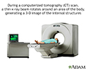
CT stands for computerized tomography. In this procedure, a thin X-ray beam is rotated around the area of the body to be visualized. Using very complicated mathematical processes called algorithms, the computer is able to generate a 3-D image of a section through the body. CT scans are very detailed and provide excellent information for the physician.
CT scan
illustration
Digestive system - illustration
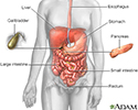
The esophagus, stomach, large and small intestine, aided by the liver, gallbladder and pancreas convert the nutritive components of food into energy and break down the non-nutritive components into waste to be excreted.
Digestive system
illustration
Liver cirrhosis - CT scan - illustration
A CT scan of the upper abdomen showing cirrhosis of the liver.
Liver cirrhosis - CT scan
illustration
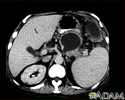
Liver metastases, CT scan - illustration
A CT scan of the upper abdomen showing multiple metastasis (cancer that has spread) in the liver of a patient with carcinoma of the large bowel. Note the dark areas in the liver (left side and center of picture).
Liver metastases, CT scan
illustration
Lymph node metastases, CT scan - illustration
A CT scan of the middle abdomen showing a large tumor mass due to metastasis (spreading cancer) in abdominal lymph nodes.
Lymph node metastases, CT scan
illustration
Lymphoma, malignant - CT scan - illustration
This abdominal CT scan shows tumor masses (malignant lymphomas) in the area behind the peritoneal cavity (retroperitoneal space).
Lymphoma, malignant - CT scan
illustration
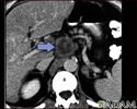
Pancreatic cancer, CT scan - illustration
A CT scan of the upper abdomen showing a tumor (pancreas carcinoma) in the head of the pancreas, seen here in the middle of the picture.
Pancreatic cancer, CT scan
illustration
Pancreatic pseudocyst - CT scan - illustration
A CT scan of the upper abdomen showing a pseudocyst in the corpus, or tail, of the pancreas.
Pancreatic pseudocyst - CT scan
illustration
Peritoneal and ovarian cancer, CT scan - illustration
A CT scan series of the lower abdomen showing ovarian cancer that has metastasized (spread) to the peritoneum.
Peritoneal and ovarian cancer, CT scan
illustration
Spleen metastasis - CT scan - illustration
This CT scan of the upper abdomen shows multiple tumors in the liver and spleen that have spread (metastasized) from an original intestinal cancer (carcinoma).
Spleen metastasis - CT scan
illustration
Normal external abdomen - illustration
The abdomen is the area of the body between the chest and pelvis. Some of the large internal organs comprised in this area are the liver, stomach, kidneys, and intestines.
Normal external abdomen
illustration


CT scan - illustration
CT stands for computerized tomography. In this procedure, a thin X-ray beam is rotated around the area of the body to be visualized. Using very complicated mathematical processes called algorithms, the computer is able to generate a 3-D image of a section through the body. CT scans are very detailed and provide excellent information for the physician.
CT scan
illustration
Digestive system - illustration
The esophagus, stomach, large and small intestine, aided by the liver, gallbladder and pancreas convert the nutritive components of food into energy and break down the non-nutritive components into waste to be excreted.
Digestive system
illustration
Liver cirrhosis - CT scan - illustration
A CT scan of the upper abdomen showing cirrhosis of the liver.
Liver cirrhosis - CT scan
illustration
Liver metastases, CT scan - illustration
A CT scan of the upper abdomen showing multiple metastasis (cancer that has spread) in the liver of a patient with carcinoma of the large bowel. Note the dark areas in the liver (left side and center of picture).
Liver metastases, CT scan
illustration
Lymph node metastases, CT scan - illustration
A CT scan of the middle abdomen showing a large tumor mass due to metastasis (spreading cancer) in abdominal lymph nodes.
Lymph node metastases, CT scan
illustration
Lymphoma, malignant - CT scan - illustration
This abdominal CT scan shows tumor masses (malignant lymphomas) in the area behind the peritoneal cavity (retroperitoneal space).
Lymphoma, malignant - CT scan
illustration
Pancreatic cancer, CT scan - illustration
A CT scan of the upper abdomen showing a tumor (pancreas carcinoma) in the head of the pancreas, seen here in the middle of the picture.
Pancreatic cancer, CT scan
illustration
Pancreatic pseudocyst - CT scan - illustration
A CT scan of the upper abdomen showing a pseudocyst in the corpus, or tail, of the pancreas.
Pancreatic pseudocyst - CT scan
illustration
Peritoneal and ovarian cancer, CT scan - illustration
A CT scan series of the lower abdomen showing ovarian cancer that has metastasized (spread) to the peritoneum.
Peritoneal and ovarian cancer, CT scan
illustration
Spleen metastasis - CT scan - illustration
This CT scan of the upper abdomen shows multiple tumors in the liver and spleen that have spread (metastasized) from an original intestinal cancer (carcinoma).
Spleen metastasis - CT scan
illustration
Normal external abdomen - illustration
The abdomen is the area of the body between the chest and pelvis. Some of the large internal organs comprised in this area are the liver, stomach, kidneys, and intestines.
Normal external abdomen
illustration
Review Date: 7/15/2024
Reviewed By: Jason Levy, MD, FSIR, Northside Radiology Associates, Atlanta, GA. Also reviewed by David C. Dugdale, MD, Medical Director, Brenda Conaway, Editorial Director, and the A.D.A.M. Editorial team.
 All rights reserved.