X-ray
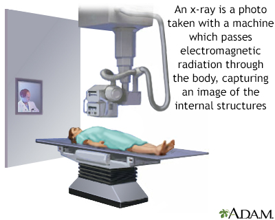
X-rays are a type of electromagnetic radiation, just like visible light.
An x-ray machine sends individual x-ray waves through the body. The images are recorded on a computer or film.
- Structures that are dense (such as bone) will block most of the x-ray, and will appear white.
- Metal and contrast media (special dye used to highlight areas of the body) will also appear white.
- Structures containing air will be black, and muscle, fat, and fluid will appear as shades of gray.
How the Test is Performed
The test is done in a hospital radiology department or in your health care provider's office. How you are positioned depends on the type of x-ray being done. Several different x-ray views may be needed.
You need to stay still when you are having an x-ray. Motion can cause blurry images. You may be asked to hold your breath or not move for a second or two when the image is being taken.
The following are common types of x-rays:
- Abdominal x-ray
Abdominal x-ray
An abdominal x-ray is an imaging test to look at organs and structures in the abdomen. Organs include the liver, spleen, stomach, and intestines. T...
 ImageRead Article Now Book Mark Article
 - Barium x-ray
Barium x-ray
Barium enema is a special x-ray of the large intestine, which includes the colon and rectum.
 ImageRead Article Now Book Mark Article
 - Bone x-ray
Bone x-ray
A bone x-ray is an imaging test to look at the bones.
 ImageRead Article Now Book Mark Article
 - Chest x-ray
Chest x-ray
A chest x-ray is an x-ray of the chest, lungs, heart, large arteries, ribs, and diaphragm.
 ImageRead Article Now Book Mark Article
 - Dental x-ray
Dental x-ray
Dental x-rays are a type of image of the teeth and mouth. X-rays are a form of high energy electromagnetic radiation. The x-rays penetrate the body...
 ImageRead Article Now Book Mark Article
 - Extremity x-ray
Extremity x-ray
An extremity x-ray is an image of the hands, wrist, feet, ankle, leg, thigh, forearm humerus or upper arm, hip, shoulder or all of these areas. The ...
ImageRead Article Now Book Mark Article - Hand x-ray
- Joint x-ray
Joint x-ray
This test is an x-ray of a knee, shoulder, hip, wrist, ankle, or other joint.
 ImageRead Article Now Book Mark Article
 - Lumbosacral spine x-ray
Lumbosacral spine x-ray
A lumbosacral spine x-ray is a picture of the small bones (vertebrae) in the lower part of the spine. This area includes the lumbar region and the s...
 ImageRead Article Now Book Mark Article
 - Neck x-ray
Neck x-ray
A neck x-ray is an imaging test to look at the cervical vertebrae. These are the 7 bones of the spine in the neck.
ImageRead Article Now Book Mark Article - Pelvis x-ray
Pelvis x-ray
A pelvis x-ray is a picture of the bones in and around both hips. The pelvis connects the legs to the body.
 ImageRead Article Now Book Mark Article
 - Sinus x-ray
Sinus x-ray
A sinus x-ray is an imaging test to look at the sinuses. These are the air-filled spaces in the front of the skull.
 ImageRead Article Now Book Mark Article
 - Skull x-ray
Skull x-ray
A skull x-ray is a picture of the bones surrounding the brain, including the facial bones, the nose, and the sinuses.
ImageRead Article Now Book Mark Article - Thoracic spine x-ray
Thoracic spine x-ray
A thoracic spine x-ray is an x-ray of the 12 chest (thoracic) bones (vertebrae) of the spine. The vertebrae are separated by flat pads of cartilage ...
ImageRead Article Now Book Mark Article - Upper GI and small bowel series
Upper GI and small bowel series
An upper GI and small bowel series is a set of x-rays taken to examine the esophagus, stomach, and small intestine. Barium enema is a different test ...
 ImageRead Article Now Book Mark Article
 - X-ray of the skeleton
X-ray of the skeleton
A skeletal x-ray is an imaging test used to look at your bones. It is used to detect fractures, tumors, or conditions that cause wearing away (degen...
ImageRead Article Now Book Mark Article
How to Prepare for the Test
Before the x-ray, tell your health care team if you are pregnant or may be pregnant.
You will need to remove all jewelry. Metal can cause unclear images. You may need to wear a hospital gown.
How the Test will Feel
X-rays are painless. Some body positions needed during an x-ray may be uncomfortable for a short time.
Risks
X-rays are monitored and regulated so you get the minimum amount of radiation exposure needed to produce the image.
For most x-rays, your risk for cancer, or if you are pregnant, the risk for birth defects in your unborn baby is very low. Most experts feel that the benefits of appropriate x-ray imaging greatly outweigh any risks.
Young children and babies in the womb are more sensitive to the risks of x-rays. Tell your provider if you think you might be pregnant.
Reviewed By
Jason Levy, MD, FSIR, Northside Radiology Associates, Atlanta, GA. Also reviewed by David C. Dugdale, MD, Medical Director, Brenda Conaway, Editorial Director, and the A.D.A.M. Editorial team.
Mettler FA. Introduction: an approach to image interpretation. In: Mettler FA, ed. Essentials of Radiology. 4th ed. Philadelphia, PA: Elsevier; 2019:chap 1.
Rodney WM, Rodney JRM, Arnold KMR. Principles of x-ray interpretation. In: Fowler GC, ed. Pfenninger and Fowler's Procedures for Primary Care. 4th ed. Philadelphia, PA: Elsevier; 2020:chap 235.

 All rights reserved.