Abdominal mass
Mass in the abdomenAn abdominal mass is swelling in one part of the belly area (abdomen).
Considerations
An abdominal mass is often found during a routine physical exam. Most of the time, the mass develops slowly. You may not be able to feel the mass.
Physical exam
During a physical examination, a health care provider checks your body to determine if you do or do not have a physical problem. A physical examinati...

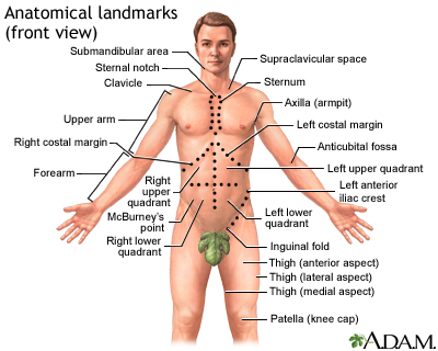
Locating the mass helps your health care provider make a diagnosis. For example, the abdomen can be divided into four areas:
- Right-upper quadrant
- Left-upper quadrant
- Right-lower quadrant
- Left-lower quadrant
Other terms used to describe the location of abdominal pain or masses include:
- Epigastric -- center of the abdomen just below the rib cage
- Periumbilical -- area around the belly button
The location of the mass and its firmness, texture, and other qualities can provide clues to its cause.
Causes
Several conditions can cause an abdominal mass:
- Abdominal aortic aneurysm can cause a pulsating mass around the navel.
Abdominal aortic aneurysm
The aorta is the main blood vessel that supplies blood to the abdomen, pelvis, and legs. An abdominal aortic aneurysm (AAA) occurs when an area of t...
 ImageRead Article Now Book Mark Article
 - Bladder distention (urinary bladder over-filled with fluid) can cause a firm mass in the center of the lower abdomen above the pelvic bones. In extreme cases, it can reach as far up as the navel.
Bladder distention
Urge incontinence occurs when you have a strong, sudden need to urinate that is difficult to delay. The bladder then squeezes, or spasms, and you ma...
 ImageRead Article Now Book Mark Article
 - Cholecystitis can cause a very tender mass that is felt below the liver in the right-upper quadrant (occasionally).
Cholecystitis
Acute cholecystitis is sudden swelling and irritation of the gallbladder. It causes severe belly pain.
 ImageRead Article Now Book Mark Article
 - Colon cancer can cause a mass almost anywhere in the abdomen.
Colon cancer
Colorectal cancer is cancer that starts in the large intestine (colon) or the rectum (end of the colon). It is also sometimes simply called colon ca...
 ImageRead Article Now Book Mark Article
 - Crohn disease or bowel obstruction can cause many tender, sausage-shaped masses anywhere in the abdomen.
Crohn disease
Crohn disease is a disease where parts of the digestive tract become inflamed. It most often involves the lower end of the small intestine and the be...
 ImageRead Article Now Book Mark Article
 - Diverticulitis can cause a mass that is usually located in the left-lower quadrant.
Diverticulitis
Diverticula are small, bulging sacs or pouches that form on the inner wall of the intestine. Diverticulitis occurs when these pouches become inflame...
 ImageRead Article Now Book Mark Article
 - Gallbladder tumor can cause a tender, irregularly shaped mass in the right-upper quadrant.
- Hydronephrosis (fluid-filled kidney) can cause a smooth, spongy-feeling mass in one or both sides or toward the back (flank area).
Hydronephrosis
Hydronephrosis is swelling of one kidney due to a backup of urine. This problem may occur in one kidney.
 ImageRead Article Now Book Mark Article
 - Kidney cancer can sometimes cause a smooth, firm, but not tender mass in the abdomen.
Kidney cancer
Renal cell carcinoma is a type of kidney cancer that starts in the lining of very small tubes (tubules) in the kidney.
 ImageRead Article Now Book Mark Article
 - Liver cancer can cause a firm, lumpy mass in the right upper quadrant.
Liver cancer
Hepatocellular carcinoma is cancer that starts in the liver.
 ImageRead Article Now Book Mark Article
 - Liver enlargement (hepatomegaly) can cause a firm, irregular mass below the right rib cage, or on the left side in the stomach area.
Liver enlargement
Enlarged liver refers to swelling of the liver beyond its normal size. Hepatomegaly is another word to describe this problem. If both the liver and ...
 ImageRead Article Now Book Mark Article
 - Neuroblastoma, a cancerous tumor often found in the lower abdomen can cause a mass (this cancer mainly occurs in children and infants).
Neuroblastoma
Neuroblastoma is a very rare type of cancerous tumor that develops from nerve tissue. It usually occurs in infants and children.
 ImageRead Article Now Book Mark Article
 - Ovarian cyst can cause a smooth, rounded, rubbery mass above the pelvis in the lower abdomen.
Ovarian cyst
An ovarian cyst is a sac filled with fluid that forms on or inside an ovary. This article is about cysts that form during your monthly menstrual cycl...
 ImageRead Article Now Book Mark Article
 - Pancreatic abscess can cause a mass in the upper abdomen in the epigastric area.
Pancreatic abscess
A pancreatic abscess is an area filled with pus within the pancreas.
 ImageRead Article Now Book Mark Article
 - Pancreatic pseudocyst can cause a lumpy mass in the upper abdomen in the epigastric area.
Pancreatic pseudocyst
A pancreatic pseudocyst is a fluid-filled sac in the abdomen that arises from the pancreas. It may also contain tissue from the pancreas, enzymes, a...
 ImageRead Article Now Book Mark Article
 - Pregnancy causes a mass above the pelvis in the lower abdomen that may be mistaken for an illness.
- Spleen enlargement (splenomegaly) can sometimes be felt in the left-upper quadrant.
Spleen enlargement
Splenomegaly is a larger-than-normal spleen. The spleen is an organ in the upper left part of the belly.
 ImageRead Article Now Book Mark Article
 - Stomach cancer can cause a mass in the left-upper abdomen in the stomach area (epigastric) if the cancer is large.
Stomach cancer
Stomach cancer is cancer that starts in the stomach.
 ImageRead Article Now Book Mark Article
Cancer
Cancer is the uncontrolled growth of abnormal cells in the body. Cancerous cells are also called malignant cells.
Read Article Now Book Mark Article - Uterine leiomyoma (fibroids) can cause a round, lumpy mass above the pelvis in the lower abdomen (sometimes can be felt if the fibroids are large).
Leiomyoma
Uterine fibroids are tumors that grow in a woman's womb (uterus). These growths are typically not cancerous (benign), and do not become cancerous....
 ImageRead Article Now Book Mark Article
 - Volvulus can cause a mass anywhere in the abdomen.
Volvulus
A volvulus is a twisting of the intestine that can occur in childhood. It causes a blockage that may cut off blood flow. Part of the intestine may ...
 ImageRead Article Now Book Mark Article
 - Ureteropelvic junction obstruction can cause a mass in the lower abdomen.
Ureteropelvic junction obstruction
Ureteropelvic junction (UPJ) obstruction is a blockage at the point where part of the kidney attaches to one of the tubes to the bladder (ureters). ...
 ImageRead Article Now Book Mark Article
Home Care
All abdominal masses should be examined as soon as possible by the provider.
Changing your body position may help relieve pain due to an abdominal mass.
When to Contact a Medical Professional
Get medical help right away if you have a pulsating lump in your abdomen along with severe abdominal pain. This could be a sign of a ruptured aortic aneurysm, which is an emergency condition.
Abdominal pain
Abdominal pain is pain that you feel anywhere between your chest and groin. This is often referred to as the stomach region or belly.

Contact your provider if you notice any type of abdominal mass.
What to Expect at Your Office Visit
In nonemergency situations, your provider will perform a physical exam and ask questions about your symptoms and medical history.
In an emergency situation, you will be stabilized first. Then, your provider will examine your abdomen and ask questions about your symptoms and medical history, such as:
- Where is the mass located?
- When did you notice the mass?
- Does it come and go?
- Has the mass changed in size or position? Has it become more or less painful?
- What other symptoms do you have?
- Is it possible that you are pregnant?
A pelvic or rectal exam may be needed in some cases. Tests that may be done to find the cause of an abdominal mass include:
- Abdominal and pelvic CT scan
Abdominal and pelvic CT scan
An abdominal CT scan is an imaging test that uses x-rays to create cross-sectional pictures of the belly area. CT stands for computed tomography....
 ImageRead Article Now Book Mark Article
 - Abdominal and pelvic ultrasound
Abdominal and pelvic ultrasound
Abdominal ultrasound is a type of imaging test. It is used to look at organs in the abdomen, including the liver, gallbladder, pancreas, and kidneys...
 ImageRead Article Now Book Mark Article
 - Angiography
Angiography
An arteriogram is an imaging test that uses x-rays and a special dye to see inside the arteries. It can be used to view arteries in the heart, brain...
 ImageRead Article Now Book Mark Article
 - Barium enema
Barium enema
Barium enema is a special x-ray of the large intestine, which includes the colon and rectum.
 ImageRead Article Now Book Mark Article
 - Blood tests
- Colonoscopy
Colonoscopy
A colonoscopy is an exam that views the inside of the colon (large intestine) and rectum, using a tool called a colonoscope. The colonoscope has a sm...
 ImageRead Article Now Book Mark Article
 - Esophagogastroduodenoscopy (EGD)
Esophagogastroduodenoscopy
Esophagogastroduodenoscopy (EGD) is a test to examine the lining of the esophagus, stomach, and first part of the small intestine (the duodenum)....
 ImageRead Article Now Book Mark Article
 - Paracentesis
- Sigmoidoscopy
Sigmoidoscopy
Sigmoidoscopy using a flexible scope is a procedure used to see inside the sigmoid colon and rectum. The sigmoid colon is the area of the large inte...
 ImageRead Article Now Book Mark Article
 - Stool analysis
- Urine tests
- X-rays of the chest or abdomen
Chest
A chest x-ray is an x-ray of the chest, lungs, heart, large arteries, ribs, and diaphragm.
 ImageRead Article Now Book Mark Article
Abdomen
An abdominal x-ray is an imaging test to look at organs and structures in the abdomen. Organs include the liver, spleen, stomach, and intestines. T...
 ImageRead Article Now Book Mark Article
References
Ball JW, Dains JE, Flynn JA, Solomon BS, Stewart RW. Abdomen. In: Ball JW, Dains JE, Flynn JA, Solomon BS, Stewart RW, eds. Seidel's Guide to Physical Examination. 10th ed. St Louis, MO: Elsevier; 2023:chap 18.
Landmann A, Bonds M, Postier R. Acute abdomen. In: Townsend CM Jr, Beauchamp RD, Evers BM, Mattox KL, eds. Sabiston Textbook of Surgery. 21st ed. St Louis, MO: Elsevier; 2022:chap 46.
McQuaid KR. Approach to the patient with gastrointestinal disease. In: Goldman L, Cooney KA, eds. Goldman-Cecil Medicine. 27th ed. Philadelphia, PA: Elsevier; 2024:chap 118.
Anatomical landmarks adult - front - illustration
There are three body views (front, back, and side) that can help you to identify a specific body area. The labels show areas of the body which are identified either by anatomical or by common names. For example, the back of the knee is called the “popliteal fossa,” while the “flank” is an area on the side of the body.
Anatomical landmarks adult - front
illustration
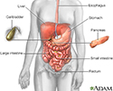
Digestive system - illustration
The esophagus, stomach, large and small intestine, aided by the liver, gallbladder and pancreas convert the nutritive components of food into energy and break down the non-nutritive components into waste to be excreted.
Digestive system
illustration
Fibroid tumors - illustration
Fibroid tumors may not need to be removed if they are not causing pain, bleeding excessively, or growing rapidly.
Fibroid tumors
illustration
Aortic aneurysm - illustration
Abdominal aortic aneurysm involves a widening, stretching, or ballooning of the aorta. There are several causes of abdominal aortic aneurysm, but the most common results from atherosclerotic disease. As the aorta gets progressively larger over time there is increased chance of rupture.
Aortic aneurysm
illustration
- Osteoporosis(Alt. Medicine)
- Obesity(Alt. Medicine)
- Diverticular disease(Alt. Medicine)
- Crohn disease(Alt. Medicine)
- Weight control and diet - InDepth(In-Depth)
- Ulcerative colitis(Alt. Medicine)
- Ovarian cancer - InDepth(In-Depth)
- Cystic fibrosis(Alt. Medicine)
- Constipation(Alt. Medicine)
- Peptic ulcer(Alt. Medicine)
Review Date: 10/9/2024
Reviewed By: Linda J. Vorvick, MD, Clinical Professor, Department of Family Medicine, UW Medicine, School of Medicine, University of Washington, Seattle, WA. Also reviewed by David C. Dugdale, MD, Medical Director, Brenda Conaway, Editorial Director, and the A.D.A.M. Editorial team.


