
Photophobia
Light sensitivity; Vision - light sensitive; Eyes - sensitivity to lightPhotophobia is eye discomfort in bright light.
Considerations
Photophobia is common. For many people, the problem is not due to any disease. Severe photophobia may occur with eye problems. It can cause bad eye pain, even in low light.
Causes
Causes may include:
- Acute iritis or uveitis (inflammation inside the eye)
Iritis
Uveitis is swelling and inflammation of the uvea. The uvea is the middle layer of the wall of the eye. The uvea supplies blood for the iris at the ...
 ImageRead Article Now Book Mark Article
Uveitis
Uveitis is swelling and inflammation of the uvea. The uvea is the middle layer of the wall of the eye. The uvea supplies blood for the iris at the ...
ImageRead Article Now Book Mark Article - Burns to the eye
- Corneal abrasion
Corneal abrasion
Corneal injury is a wound to the part of the eye known as the cornea. The cornea is the crystal clear (transparent) tissue that covers the front of ...
 ImageRead Article Now Book Mark Article
 - Corneal ulcer
Corneal ulcer
The cornea is the clear tissue at the front of the eye. A corneal ulcer is an open sore in the outer layer of the cornea. It is often caused by inf...
ImageRead Article Now Book Mark Article - Drugs such as amphetamines, atropine, cocaine, cyclopentolate, idoxuridine, phenylephrine, scopolamine, trifluridine, tropicamide, and vidarabine
Cocaine
A toxicology screen refers to various tests that determine the type and approximate amount of legal and illegal drugs a person has taken by measuring...
 ImageRead Article Now Book Mark Article
 - Excessive wearing of contact lenses, or wearing poorly-fitting contact lenses
- Eye disease, injury, or infection (such as chalazion, episcleritis, glaucoma)
Chalazion
A chalazion is a small bump in the eyelid caused by a blockage of a tiny oil gland.
ImageRead Article Now Book Mark ArticleEpiscleritis
Episcleritis is irritation and inflammation of the episclera, a thin layer of tissue covering the white part (sclera) of the eye. It is not an infec...
 ImageRead Article Now Book Mark Article
Glaucoma
Glaucoma is a group of eye conditions that can damage the optic nerve. This nerve sends the images you see to your brain. Most often, optic nerve da...
 ImageRead Article Now Book Mark Article
 - Eye testing when the eyes have been dilated
- Meningitis
Meningitis
Meningitis is an infection of the membranes covering the brain and spinal cord. This covering is called the meninges.
 ImageRead Article Now Book Mark Article
 - Migraine headache
- Recovery from eye surgery
Home Care
Things you can do to ease light sensitivity include:
- Avoid sunlight
- Close your eyes
- Wear dark glasses
- Darken the room
If eye pain is severe, see your health care provider about the cause of light sensitivity. Proper treatment may cure the problem. Get medical help right away if your pain is moderate to severe, even in low-light conditions.
When to Contact a Medical Professional
Call your provider if:
- Light sensitivity is severe or painful. (For example, you need to wear sunglasses indoors.)
- Sensitivity occurs with headaches, red eye or blurred vision or does not go away in a day or two.
Red eye
Eye redness is most often due to swollen or dilated blood vessels. This makes the surface of the eye look red or bloodshot.
 ImageRead Article Now Book Mark Article
Blurred vision
There are many types of eye problems and vision disturbances, such as: Halos Blurred vision (the loss of sharpness of vision and the inability to see...
 ImageRead Article Now Book Mark Article
What to Expect at Your Office Visit
The provider will perform a physical exam, including an eye exam. You may be asked the following questions:
Physical exam
During a physical examination, a health care provider checks your body to determine if you do or do not have a physical problem. A physical examinati...

- When did the light sensitivity begin?
- How bad is the pain? Does it hurt all the time or just sometimes?
- Do you need to wear dark glasses or stay in dark rooms?
- Did a provider recently dilate your pupils?
- What medicines do you take? Have you used any eye drops?
- Do you use contact lenses?
- Have you used soaps, lotions, cosmetics, or other chemicals around your eyes?
- Does anything make the sensitivity better or worse?
- Have you been injured?
- What other symptoms do you have?
Tell your provider if you have any of these symptoms:
- Pain in the eye
Pain in the eye
Pain in the eye may be described as a burning, throbbing, aching, or stabbing sensation in or around the eye. It may also feel like you have a forei...
ImageRead Article Now Book Mark Article - Nausea or dizziness
- Headache or neck stiffness
- Blurred vision
Blurred vision
There are many types of eye problems and vision disturbances, such as: Halos Blurred vision (the loss of sharpness of vision and the inability to see...
ImageRead Article Now Book Mark Article - Sore or wound in eye
- Redness, itching, or swelling
- Numbness or tingling elsewhere in the body
- Changes in hearing
The following tests may be done:
- Corneal scraping
- Lumbar puncture (most often done by a neurologist)
Lumbar puncture
Cerebrospinal fluid (CSF) collection is a test to look at the fluid that surrounds the brain and spinal cord. CSF acts as a cushion, protecting the b...
 ImageRead Article Now Book Mark Article
 - Pupil dilation
- Pupillary response to light
- Slit-lamp exam
Slit-lamp exam
The slit-lamp examination looks at structures that are at the front of the eye.
ImageRead Article Now Book Mark Article
References
Datoo O'Keefe GA. Idiopathic and other anterior uveitis syndromes. In: Yanoff M, Duker JS, eds. Ophthalmology. 6th ed. Philadelphia, PA: Elsevier; 2023:chap 7.19.
Ghanem RC, Ghanem MA, Azar DT. LASIK complications and their management. In: Azar DT, ed. Refractive Surgery. 3rd ed. Philadelphia, PA: Elsevier; 2020:chap 15.
Olson JA. Medical ophthalmology. In: Penman ID, Ralston SH, Strachan MWJ, Hobson RP, eds. Davidson's Principles and Practice of Medicine. 24th ed. Philadelphia, PA: Elsevier; 2023:chap 30.
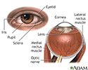
External and internal eye anatomy - illustration
The cornea allows light to enter the eye. As light passes through the eye the iris changes shape by expanding and letting more light through or constricting and letting less light through to change pupil size. The lens then changes shape to allow the accurate focusing of light on the retina. Light excites photoreceptors that eventually, through a chemical process, transmit nerve signals through the optic nerve to the brain. The brain processes these nerve impulses into sight.
External and internal eye anatomy
illustration
Review Date: 5/10/2023
Reviewed By: Franklin W. Lusby, MD, Ophthalmologist, Lusby Vision Institute, La Jolla, CA. Also reviewed by David C. Dugdale, MD, Medical Director, Brenda Conaway, Editorial Director, and the A.D.A.M. Editorial team.
 All rights reserved.