
Nystagmus
Back and forth eye movements; Involuntary eye movements; Rapid eye movements from side to side; Uncontrolled eye movements; Eye movements - uncontrollableNystagmus is a term to describe uncontrollable movements of the eyes that may be:
- Side to side (horizontal nystagmus)
- Up and down (vertical nystagmus)
- Rotary (rotary or torsional nystagmus)
Depending on the cause, these movements may be in both eyes or in just one eye.
Nystagmus can affect vision, balance, and coordination.
Considerations
The involuntary eye movements of nystagmus are caused by abnormal function in the areas of the brain that control eye movements. The part of the inner ear that senses movement and position (the labyrinth) helps control eye movements.
There are two forms of nystagmus:
- Infantile nystagmus syndrome (INS) is present at birth (congenital).
- Acquired nystagmus develops later in life because of a disease or injury.
Causes
NYSTAGMUS THAT IS PRESENT AT BIRTH (infantile nystagmus syndrome, or INS)
INS is usually mild. It does not become more severe, and it is not related to any other disorder.
People with this condition are usually not aware of the eye movements, but other people may see them. If the movements are large, sharpness of vision (visual acuity) may be less than 20/20. Surgery may improve vision.
Nystagmus may be caused by congenital diseases of the eye. Although this is rare, an eye doctor (ophthalmologist) should evaluate any child with nystagmus to check for eye disease.
ACQUIRED NYSTAGMUS
The most common cause of acquired nystagmus is certain drugs or medicines. Phenytoin (Dilantin) - an antiseizure medicine, excessive alcohol, or any sedating medicine can impair the labyrinth's function.
Other causes include:
-
Inner ear disorders such as benign positional vertigo, labyrinthitis or Meniere disease
Benign positional vertigo
Benign positional vertigo is the most common type of vertigo. Vertigo is the feeling that you are spinning or that everything is spinning around you...
 ImageRead Article Now Book Mark Article
Labyrinthitis
Labyrinthitis is irritation and swelling of the inner ear. It can cause vertigo and hearing loss.
 ImageRead Article Now Book Mark Article
Meniere disease
Ménière disease is an inner ear disorder that affects balance and hearing.
ImageRead Article Now Book Mark Article - Head injury from motor vehicle accidents
- Stroke
Stroke
A stroke occurs when blood flow to a part of the brain stops. A stroke is sometimes called a "brain attack. " If blood flow is cut off for longer th...
 ImageRead Article Now Book Mark Article
 - Infections such as Lyme disease and syphilis
-
Thiamine or vitamin B12 deficiency
Vitamin B12
Vitamin B12 is a water-soluble vitamin. Water-soluble vitamins dissolve in water. After the body uses what it needs of these vitamins, leftover amo...
 ImageRead Article Now Book Mark Article
 - Multiple sclerosis
- Brain tumors
- Autoimmune disease
- Paraneoplastic syndromes (due to cancer elsewhere in the body)
- Seizures
Nystagmus can also be a symptom of other neurological disorders.
Home Care
You may need to make changes in the home to help with dizziness, visual problems, or nervous system disorders.
Dizziness
Dizziness is a term that is often used to describe 2 different symptoms: lightheadedness and vertigo. Lightheadedness is a feeling that you might fai...

Visual problems
There are many types of eye problems and vision disturbances, such as: Halos Blurred vision (the loss of sharpness of vision and the inability to see...

When to Contact a Medical Professional
Contact your health care provider if you have symptoms of nystagmus or think you might have this condition.
What to Expect at Your Office Visit
Your provider will take a careful history and perform a thorough physical examination, focusing on the nervous system and inner ear. The provider may ask you to wear a pair of goggles that magnify your eyes for part of the examination.
To check for nystagmus, the provider may use the following procedure:
- You spin around for about 30 seconds, stop, and try to stare at an object.
- Your eyes will first move slowly in one direction, then will move quickly in the opposite direction.
If you have nystagmus due to a medical condition, the type and severity of the eye movements during this maneuver will depend on the cause.
You may have the following tests:
- CT scan of the head
CT scan
A computed tomography (CT) scan is an imaging method that uses x-rays to create pictures of cross-sections of the body. Related tests include:Abdomin...
 ImageRead Article Now Book Mark Article
 - Electro-oculography: An electrical method of measuring eye movements using tiny electrodes
- MRI of the head
MRI of the head
A head MRI (magnetic resonance imaging) is an imaging test that uses powerful magnets and radio waves to create pictures of the brain and surrounding...
ImageRead Article Now Book Mark Article - Electronystagmography: Vestibular testing by recording the movements of the eyes
Electronystagmography
Electronystagmography is a test that looks at eye movements to see how well nerves in the brain are working. These nerves are:Vestibular nerve (eigh...
Read Article Now Book Mark Article
There is no treatment for most cases of congenital nystagmus. Treatment for acquired nystagmus depends on the cause. In some cases, nystagmus cannot be reversed. In cases due to medicines or infection, the nystagmus usually goes away after the cause has gotten better.
Some treatments may help improve the visual function of people with infantile nystagmus syndrome:
- Prisms
- Surgery such as tenotomy
- Drug therapies for infantile nystagmus
References
Olitsky SE, Marsh JD. Disorders of eye movement and alignment. In: Kliegman RM, St. Geme JW, Blum NJ, Shah SS, Tasker RC, Wilson KM, eds. Nelson Textbook of Pediatrics. 21st ed. Philadelphia, PA: Elsevier; 2020:chap 641.
Quiros PA, Chang MY. Nystagmus, saccadic intrusions, and oscillations. In: Yanoff M, Duker JS, eds. Ophthalmology. 6th ed. Philadelphia, PA: Elsevier; 2023:chap 9.19.
Rucker JC, Lavin PJM. Neuro-ophthalmology: ocular motor system. In: Jankovic J, Mazziotta JC, Pomeroy SL, Newman NJ, eds. Bradley and Daroff's Neurology in Clinical Practice. 8th ed. Philadelphia, PA: Elsevier; 2022:chap 18.
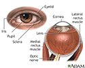
External and internal eye anatomy - illustration
The cornea allows light to enter the eye. As light passes through the eye the iris changes shape by expanding and letting more light through or constricting and letting less light through to change pupil size. The lens then changes shape to allow the accurate focusing of light on the retina. Light excites photoreceptors that eventually, through a chemical process, transmit nerve signals through the optic nerve to the brain. The brain processes these nerve impulses into sight.
External and internal eye anatomy
illustration
Review Date: 1/23/2023
Reviewed By: Joseph V. Campellone, MD, Department of Neurology, Cooper Medical School of Rowan University, Camden, NJ. Review provided by VeriMed Healthcare Network. Also reviewed by David C. Dugdale, MD, Medical Director, Brenda Conaway, Editorial Director, and the A.D.A.M. Editorial team.
 All rights reserved.