Vision problems
Vision impairment; Impaired vision; Blurred visionThere are many types of eye problems and vision disturbances, such as:
- Halos
- Blurred vision (the loss of sharpness of vision and the inability to see fine details)
- Blind spots or scotomas (dark "holes" in the vision in which nothing can be seen)
Vision loss and blindness are the most severe vision problems.
Considerations
Regular eye checkups from an ophthalmologist or optometrist are important. They should be done once a year if you are over age 65 years or older. Some experts recommend annual eye exams starting at an earlier age.
How long you go between exams is based on how long you can wait before detecting an eye problem that has no symptoms. Your health care provider will recommend earlier and more frequent exams if you have known eye problems or conditions that are known to cause eye problems. These include diabetes or high blood pressure.
These important steps can prevent eye and vision problems:
- Wear sunglasses to protect your eyes.
- Wear safety glasses when hammering, grinding, or using power tools.
- If you need glasses or contact lenses, keep the prescription up to date.
- Do not smoke.
- Limit how much alcohol you drink.
- Stay at a healthy weight.
- Keep your blood pressure and cholesterol under control.
- Keep your blood sugar under control if you have diabetes.
- Eat foods rich in antioxidants, like green leafy vegetables.
Causes
Vision changes and problems can be caused by many different conditions. Some include:
- Presbyopia -- Difficulty focusing on objects that are close. This problem often becomes noticeable in your early to mid-40s and is very common.
Presbyopia
Presbyopia is a condition in which the lens of the eye loses its ability to focus. This makes it hard to see objects up close.
 ImageRead Article Now Book Mark Article
 - Cataracts -- Cloudiness of the eye lens, causing poor nighttime vision, halos around lights, and sensitivity to glare. Cataracts are common in older people.
- Glaucoma -- Increased pressure in the eye, which is most often painless. Vision will be normal at first, but over time you can develop poor night vision, blind spots, and a loss of vision to either side. Some types of glaucoma can also happen suddenly, which is a medical emergency.
Glaucoma
Glaucoma is a group of eye conditions that can damage the optic nerve. This nerve sends the images you see to your brain. Most often, optic nerve da...
 ImageRead Article Now Book Mark Article
 - Diabetic eye disease.
Diabetic eye disease
Diabetes can harm the eyes. It can damage the small blood vessels in the retina, the back part of your eye. This condition is called diabetic retin...
 ImageRead Article Now Book Mark Article
 - Macular degeneration -- Loss of central vision, blurred vision (particularly while reading), distorted vision (straight lines will appear to be wavy), and colors that look faded. This is the most common cause of blindness in people over age 60 years.
Macular degeneration
Macular degeneration is an eye disorder that slowly destroys sharp, central vision. This makes it difficult to see fine details and read. The diseas...
 ImageRead Article Now Book Mark Article
 - Eye infection, inflammation, or injury.
- Floaters -- Tiny particles drifting inside the eye, which may be a sign of retinal detachment.
Floaters
The floating specks you sometimes see in front of your eyes are not on the surface of your eyes but inside them. They are made up of bits of cell de...
 ImageRead Article Now Book Mark Article
 - Night blindness.
Night blindness
Night blindness is poor vision at night or in dim light.
 ImageRead Article Now Book Mark Article
 - Retinal detachment -- Symptoms include floaters, sparks, or flashes of light in your vision, or a sensation of a shade or curtain hanging across part of your visual field.
Retinal detachment
Retinal detachment is a separation of the light-sensitive membrane (retina) in the back of the eye from its supporting layers.
 ImageRead Article Now Book Mark Article
 - Optic neuritis -- Inflammation of the optic nerve from infection or multiple sclerosis. You may have pain when you move your eye or touch it through the eyelid.
Optic neuritis
The optic nerve carries images of what the eye sees to the brain. When this nerve become swollen or inflamed, it is called optic neuritis. It may c...
 ImageRead Article Now Book Mark Article
Multiple sclerosis
Multiple sclerosis (MS) is an autoimmune disease that affects the brain and spinal cord (central nervous system).
 ImageRead Article Now Book Mark Article
 - Stroke or transient ischemic attack (TIA).
Stroke
A stroke occurs when blood flow to a part of the brain stops. A stroke is sometimes called a "brain attack. " If blood flow is cut off for longer th...
 ImageRead Article Now Book Mark Article
Transient ischemic attack
A transient ischemic attack (TIA) occurs when blood flow to a part of the brain stops for a brief time. A person will have stroke-like symptoms for ...
 ImageRead Article Now Book Mark Article
 - Brain tumor.
Brain tumor
A brain tumor is a group (mass) of abnormal cells that grow in the brain. This article focuses on primary brain tumors in children.
 ImageRead Article Now Book Mark Article
 - Bleeding into the eye.
- Temporal arteritis -- Inflammation of an artery in the brain that supplies blood to the optic nerve.
Temporal arteritis
Giant cell arteritis (GCA) is inflammation and damage to the blood vessels that supply blood to the head, neck, upper body and arms. It is also call...
 ImageRead Article Now Book Mark Article
 - Migraine headaches -- Spots of light, halos, or zigzag patterns that appear before the start of the headache.
Migraine headaches
A migraine is a type of headache. It may occur with symptoms such as nausea, vomiting, or sensitivity to light and sound. In most people, a throbbi...
 ImageRead Article Now Book Mark Article
Medicines may also affect your vision.
Home Care
See your provider if you have any problems with your eyesight.
When to Contact a Medical Professional
Seek emergency care from a provider who is experienced in dealing with eye emergencies if:
- You experience partial or complete blindness in one or both eyes, even if it is only temporary.
Blindness
Blindness is a lack of vision. It may also refer to a loss of vision that cannot be corrected with glasses or contact lenses. Partial blindness mean...
 ImageRead Article Now Book Mark Article
 - You experience double vision, even if it is temporary.
- You have a sensation of a shade being pulled over your eyes or a curtain being drawn from the side, above, or below.
- Blind spots, halos around lights, or areas of distorted vision appear suddenly.
- You have sudden blurred vision with eye pain, particularly if the eye is also red. A red, painful eye with blurred vision is a medical emergency.
Get a complete eye exam if you have:
- Trouble seeing objects on either side.
- Difficulty seeing at night or when reading.
- Gradual loss of the sharpness of your vision.
- Difficulty telling colors apart.
- Blurred vision when trying to view objects near or far.
- Diabetes or a family history of diabetes.
- Eye itching or discharge.
- Vision changes that seem related to medicine. (Do not stop or change a medicine without talking to your provider.)
What to Expect at Your Office Visit
Your provider will check your vision, eye movements, pupils, the back of your eye (called the retina), and eye pressure. An overall medical evaluation will be done if needed.
It will be helpful to your provider if you can describe your symptoms accurately. Think about the following ahead of time:
- Has the problem affected your vision?
- Is there blurring, halos around lights, flashing lights, or blind spots?
- Do colors seem faded?
- Do you have pain?
- Are you sensitive to light?
- Do you have tearing or discharge?
- Do you have dizziness, or does it seem like the room is spinning?
- Do you have double vision?
- Is the problem in one or both eyes?
- When did this begin? Did it occur suddenly or gradually?
- Is it constant or does it come and go?
- How often does it occur? How long does it last?
- When does it occur? Evening? Morning?
- Is there anything that makes it better? Worse?
The provider will also ask you about any eye problems you have had in the past:
- Has this ever happened before?
- Have you been given eye medicines?
- Have you had eye surgery or injuries?
- Have you recently traveled out of the country?
- Are there new things you could be allergic to, such as soaps, sprays, lotions, creams, cosmetics, laundry products, curtains, sheets, carpets, paint, or pets?
The provider will also ask about your general health and family history:
- Do you have any known allergies?
- When did you last have a general checkup?
- Are you taking any medicines?
- Have you been diagnosed with any medical conditions, such as diabetes or high blood pressure?
- What kinds of eye problems do your family members have?
The following tests may be performed:
- Dilated eye exam
- Slit-lamp examination
Slit-lamp examination
The slit-lamp examination looks at structures that are at the front of the eye.
 ImageRead Article Now Book Mark Article
 - Refraction (test for glasses)
Refraction
A refraction is an eye exam that measures a person's prescription for eyeglasses or contact lenses.
 ImageRead Article Now Book Mark Article
 - Tonometry (eye pressure test)
Tonometry
Tonometry is a test to measure the pressure inside your eyes. The test is used to screen for glaucoma. It is also used to measure how well glaucoma...
 ImageRead Article Now Book Mark Article
Treatments depend on the cause. Surgery may be needed for some conditions.
References
Chou R, Bougatsos C, Jungbauer R, et al. Screening for impaired visual acuity in older adults: updated evidence report and systematic review for the US Preventive Services Task Force. JAMA. 2022;327(21):2129-2140.PMID: 35608842 pubmed.ncbi.nlm.nih.gov/35608842/.
Cioffi GA, Liebmann JM. Diseases of the visual system. In: Goldman L, Cooney KA, eds. Goldman-Cecil Medicine. 27th ed. Philadelphia, PA: Elsevier; 2024:chap 391.
Chaves-Gnecco D, Feldman HM. Developmental/behavioral pediatrics. In: Zitelli BJ, McIntire SC, Nowalk AJ, Garrison J, eds. Zitelli and Davis' Atlas of Pediatric Physical Diagnosis. 8th ed. Philadelphia, PA: Elsevier; 2023:chap 3.
Thurtell MJ, Prasad S, Tomsak RL. Neuro-ophthalmology: afferent visual system . In: Jankovic J, Mazziotta JC, Pomeroy SL, Newman NJ, eds. Bradley and Daroff's Neurology in Clinical Practice. 8th ed. Philadelphia, PA: Elsevier; 2022:chap 16.
US Preventive Services Task Force; Grossman DC, Curry SJ, et al. Vision screening in children aged 6 months to 5 years: US Preventive Services Task Force Recommendation Statement. JAMA. 2017;318(9):836-844. PMID: 28873168 pubmed.ncbi.nlm.nih.gov/28873168/.
Crossed eyes - illustration
People are very sensitive to other individuals' eye positions. By looking at another person's eye position, one can very effectively gauge where they are looking. People are also sensitive to eyes that are not looking in the same direction, which is referred to as crossed eyes (strabismus). Other more specific medical terms refer to eyes turned either outward or inward, or that are abnormally rotated. Any appearance of crossed eyes in young children should be immediately evaluated, as should recent onset of crossed eyes in an adult.
Crossed eyes
illustration
Eye - illustration
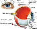
The eye is the organ of sight, a nearly spherical hollow globe filled with fluids (humors). The outer layer (sclera, or white of the eye, and cornea) is fibrous and protective. The middle layer (choroid, ciliary body and the iris) is vascular. The innermost layer (retina) is sensory nerve tissue that is light sensitive. The fluids in the eye are divided by the lens into the vitreous humor (behind the lens) and the aqueous humor (in front of the lens). The lens itself is flexible and suspended by ligaments which allow it to change shape to focus light on the retina, which is composed of sensory neurons.
Eye
illustration
Visual acuity test - illustration
Visual acuity tests may be performed in many different ways. It is a quick way to detect vision problems and is frequently used in schools or for mass screening. Driver license bureaus often use a small device that can test the eyes both together and individually.
Visual acuity test
illustration
Slit-lamp exam - illustration
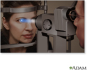
A slit-lamp, which is a specialized magnifying microscope, is used to examine the structures of the eye (including the cornea, iris, vitreous, and retina). The slit-lamp is used to examine, treat (with a laser), and photograph (with a camera) the eye.
Slit-lamp exam
illustration
Visual field test - illustration
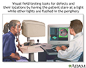
Central and peripheral vision is tested by using visual field tests. Changes may indicate eye diseases, such as glaucoma or retinitis.
Visual field test
illustration
Cataract - close-up of the eye - illustration
This photograph shows a cloudy white lens (cataract) as seen through the pupil. Cataracts are a leading cause of decreased vision in older adults, but children may have congenital cataracts. With surgery, the cataract can be removed, a new lens implanted, and the person can usually return home the same day.
Cataract - close-up of the eye
illustration
Cataract - illustration
A cataract is a cloudy or opaque area in the lens of the eye. Cataracts usually develop as a person gets older and may run in families. Other environmental factors such as smoking or exposure to toxic substances can also accelerate the development of a cataract. Cataracts can cause visual problems such as difficulty seeing at night, seeing halos around lights, and sensitivity to glare.
Cataract
illustration
Review Date: 8/5/2024
Reviewed By: Franklin W. Lusby, MD, Ophthalmologist, Lusby Vision Institute, La Jolla, CA. Also reviewed by David C. Dugdale, MD, Medical Director, Brenda Conaway, Editorial Director, and the A.D.A.M. Editorial team.


