
Eye floaters
Specks in your visionInformation
The floating specks you sometimes see in front of your eyes are not on the surface of your eyes but inside them. They are made up of bits of cell debris that drift around in the fluid (vitreous) in the back of your eye. Floaters may look like spots, specks, bubbles, threads, or clumps. Most adults have at least a few floaters. There are times when they may be more visible than at other times, such as when you are reading, especially if the floater is dense or dark.
Most of the time floaters are harmless. However, they can be a symptom of a tear in the retina. If you notice a sudden increase in floaters or if you see floaters along with flashes of light in your side vision, this may be a symptom of a retinal tear or detachment. Go to an eye doctor or emergency room if you have these symptoms.
Retina
The retina is the light-sensitive layer of tissue at the back of the eyeball. Images that come through the eye's lens are focused on the retina. Th...

Treatment
It is difficult to eliminate floaters. A vitrectomy can remove floaters, but it is a major eye operation and many doctors feel that it is too risky for a minor problem such as floaters. A YAG laser treatment can break up floaters and possibly make them less visible, but it doesn't really remove the floaters. Research is being done on femtosecond lasers (like the ones often used in LASIK) since this method may prove to be safer. Atropine eye drops, which slightly dilate the pupil, can make floaters less visible. However, the drops may cause blurred vision. Some homeopathic eye drops are marketed as treatment for floaters, but not been shown to have proven benefit.
References
Broadhead GK, Hong T, Chang AA. To treat or not to treat: management options for symptomatic vitreous floaters. Asia Pac J Ophthalmol (Phila). 2020;9(2):96-103. PMID: 32097127 pubmed.ncbi.nlm.nih.gov/32097127/.
Crouch ER, Crouch ER, Grant TR. Ophthalmology. In: Rakel RE, Rakel DP, eds. Textbook of Family Medicine. 9th ed. Philadelphia, PA: Elsevier Saunders; 2016:chap 17.
Guluma K, Lee JE. Ophthalmology. In: Walls RM, ed. Rosen's Emergency Medicine: Concepts and Clinical Practice. 10th ed. Philadelphia, PA: Elsevier; 2023:chap 57.
Sun X, Tian J, Wang J, Zhang J, Wang Y, Yuan G. Nd:YAG Laser Vitreolysis for Symptomatic Vitreous Floaters: Application of Infrared Fundus Photography in Assessing the Treatment Efficacy. J Ophthalmol. 2019; 2019:8956952. PMID: 30809388 pubmed.ncbi.nlm.nih.gov/30809388/.
Souza CE, Lima LH, Nascimento H, Zett C, Belfort R. Objective assessment of YAG laser vitreolysis in patients with symptomatic vitreous floaters. Int J Retina Vitreous. 2020;6:1. PMID: 31988795 pubmed.ncbi.nlm.nih.gov/31988795/.
Woudstra-de Jong JE, Manning-Charalampidou SS, Vingerling H, Busschbach JJ, Pesudovs K. Patient-reported outcomes in patients with vitreous floaters: a systematic literature review. Surv Ophthalmol. 2023;68(5):875-888. PMID: 37315741 pubmed.ncbi.nlm.nih.gov/37315741/.
Eye floaters - illustration
Eye floaters are small, squiggly lines or dark specks that move as if floating in your vision. They are caused by clumps of cell debris or microscopic protein fibers that drift around in the fluid that fills the back of your eye. When light enters the eye, these clumps can cast a small shadow on your retina, and this shadow is what you see as floaters.
Eye floaters
illustration
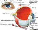
Eye - illustration
The eye is the organ of sight, a nearly spherical hollow globe filled with fluids (humors). The outer layer (sclera, or white of the eye, and cornea) is fibrous and protective. The middle layer (choroid, ciliary body and the iris) is vascular. The innermost layer (retina) is sensory nerve tissue that is light sensitive. The fluids in the eye are divided by the lens into the vitreous humor (behind the lens) and the aqueous humor (in front of the lens). The lens itself is flexible and suspended by ligaments which allow it to change shape to focus light on the retina, which is composed of sensory neurons.
Eye
illustration

Eye floaters - illustration
Eye floaters are small, squiggly lines or dark specks that move as if floating in your vision. They are caused by clumps of cell debris or microscopic protein fibers that drift around in the fluid that fills the back of your eye. When light enters the eye, these clumps can cast a small shadow on your retina, and this shadow is what you see as floaters.
Eye floaters
illustration
Eye - illustration
The eye is the organ of sight, a nearly spherical hollow globe filled with fluids (humors). The outer layer (sclera, or white of the eye, and cornea) is fibrous and protective. The middle layer (choroid, ciliary body and the iris) is vascular. The innermost layer (retina) is sensory nerve tissue that is light sensitive. The fluids in the eye are divided by the lens into the vitreous humor (behind the lens) and the aqueous humor (in front of the lens). The lens itself is flexible and suspended by ligaments which allow it to change shape to focus light on the retina, which is composed of sensory neurons.
Eye
illustration
Review Date: 11/8/2023
Reviewed By: Franklin W. Lusby, MD, Ophthalmologist, Lusby Vision Institute, La Jolla, CA. Also reviewed by David C. Dugdale, MD, Medical Director, Brenda Conaway, Editorial Director, and the A.D.A.M. Editorial team.
 All rights reserved.