Meibomianitis
Meibomian gland dysfunctionMeibomianitis is inflammation of the meibomian glands, a group of oil-releasing (sebaceous) glands in the eyelids. These glands have tiny openings to release oils onto the surface of the cornea.
Causes
Any condition that increases the oily secretions of the meibomian glands will allow excess oils to build up on the edges of the eyelids. This allows for the excess growth of bacteria that are normally present on the skin.
These problems can be caused by allergies, hormone changes during adolescence, or skin conditions such as rosacea and acne.
Rosacea
Rosacea is a chronic skin problem that makes your face turn red. It may also cause swelling and skin sores that look like acne.

Meibomianitis is often associated with blepharitis, which can cause a buildup of a dandruff-like substance at the base of the eyelashes.
Blepharitis
Blepharitis is manifest by inflamed, irritated, itchy, and reddened eyelids. It most often occurs where the eyelashes grow. Dandruff-like debris bu...

In some people with meibomianitis, the glands will be plugged so that there is less oil being made for the normal tear film. These people often have symptoms of dry eyes.
Symptoms
Symptoms include:
- Swelling and redness of eyelid edges
- Symptoms of dry eye
- Slight blurring of vision due to excess oil in tears -- most often cleared by blinking
- Frequent styes
Styes
Most bumps on the eyelid are styes. A stye is an inflamed oil gland on the edge of your eyelid, where the eyelash meets the lid. It appears as a re...
 ImageRead Article Now Book Mark Article
Exams and Tests
Meibomianitis can be diagnosed by an eye exam. Special tests are not required.
Treatment
Standard treatment involves:
- Carefully cleansing the edges of the lids
- Applying moist heat to the affected eye
These treatments will usually reduce symptoms in most cases.
Your health care provider may prescribe an antibiotic ointment to apply to the lid's edge.
Other treatments may include:
- Having an eye doctor perform meibomian gland expression to help clear the glands of secretions.
- Inserting a small tube (cannula) into each gland opening to wash out thickened oil.
- Taking a tetracycline antibiotic for several weeks.
- Using LipiFlow, a device that automatically warms the eyelid and helps clear the glands.
- Taking fish oil to improve the flow of oil from the glands.
- Using a medicine containing hypochlorous acid, that is sprayed onto the eyelids. This may be particularly useful in people who have rosacea.
You may also need treatment for general skin conditions such as acne or rosacea.
Outlook (Prognosis)
Meibomianitis is not a vision-threatening condition. However, it may be a long-term (chronic) and recurring cause of eye irritation. Many people find the treatments frustrating because results are not often immediate. Treatment, however, will often help reduce symptoms.
When to Contact a Medical Professional
Contact your provider if treatment does not lead to improvement or if styes develop.
Prevention
Keeping your eyelids clean and treating associated skin conditions will help prevent meibomianitis.
References
Foulks GN, Lemp MA. Meibomian gland dysfunction and the evaporative eye. In: Mannis MJ, Holland EJ, eds. Cornea. 5th ed. Philadelphia, PA: Elsevier; 2022:chap 33.
Hu J, Zhu S, Liu X. Efficacy and safety of a vectored thermal pulsation system (Lipiflow) in the treatment of meibomian gland dysfunction: a systematic review and meta-analysis. Graefes Arch Clin Exp Ophthalmol. 2022;260(1):25-39. PMID: 34374808 pubmed.ncbi.nlm.nih.gov/34374808/.
Starnes TW, Vasaiwala RA, Bouchard CS. Noninfectious keratitis. In: Yanoff M, Duker JS, eds. Ophthalmology. 6th ed. Philadelphia, PA: Elsevier; 2023:chap 4.17.
-
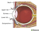
Eye anatomy - illustration
The cornea is the clear layer covering the front of the eye. The cornea works with the lens of the eye to focus images on the retina.
Eye anatomy
illustration
Review Date: 8/5/2024
Reviewed By: Franklin W. Lusby, MD, Ophthalmologist, Lusby Vision Institute, La Jolla, CA. Also reviewed by David C. Dugdale, MD, Medical Director, Brenda Conaway, Editorial Director, and the A.D.A.M. Editorial team.


