Valley fever
San Joaquin Valley fever; Coccidioidomycosis; Cocci; Desert rheumatismValley fever is an infection that occurs when the spores of the fungus Coccidioides immitis enter your body through the lungs.
Spores
A spore is a cell that certain fungi, plants (moss, ferns), and bacteria produce. Certain bacteria make spores as a way to defend themselves. Spores...
Read Article Now Book Mark ArticleCauses
Valley fever is a fungal infection most commonly seen in the desert regions of the southwestern United States, and in Central and South America. You get it by breathing in the fungus from soil. The infection starts in the lungs. It commonly affects people over 60 years of age.
Valley fever may also be called coccidioidomycosis.
Traveling to an area where the fungus is commonly seen raises your risk for this infection. However, you're more likely to develop a serious infection if you live where the fungus is found and have a weakened immune system due to:
- Anti-tumor necrosis factor (TNF) therapy
-
Cancer
Cancer
Cancer is the uncontrolled growth of abnormal cells in the body. Cancerous cells are also called malignant cells.
Read Article Now Book Mark Article -
Chemotherapy
Chemotherapy
The term chemotherapy is used to describe cancer-killing drugs. Chemotherapy may be used to:Cure the cancerShrink the cancerPrevent the cancer from ...
 ImageRead Article Now Book Mark Article
 - Glucocorticoid medicines (prednisone)
- Heart or lung conditions
-
HIV/AIDS
HIV/AIDS
Human immunodeficiency virus (HIV) is the virus that causes acquired immunodeficiency syndrome (AIDS). When a person becomes infected with HIV, the ...
 ImageRead Article Now Book Mark Article
 - Organ transplant
- Pregnancy (especially the first trimester)
People of Native American, African, or Philippine descent are disproportionately affected.
Symptoms
Most people with valley fever never have symptoms. Others may have cold- or flu-like symptoms or symptoms of pneumonia. If symptoms occur, they typically start 5 to 21 days after exposure to the fungus.
Common symptoms include:
- Ankle, feet, and leg swelling
-
Chest pain (can vary from mild to severe)
Chest pain
Chest pain is discomfort or pain that you feel anywhere along the front of your body between your neck and upper abdomen.
 ImageRead Article Now Book Mark Article
 - Cough, possibly producing blood-tinged phlegm (sputum)
- Fever and night sweats
- Headache
- Joint stiffness and pain or muscle aches
- Loss of appetite
- Painful, red lumps on lower legs (erythema nodosum)
Erythema nodosum
Erythema nodosum is an inflammatory disorder. It involves tender, red bumps (nodules) under the skin.
 ImageRead Article Now Book Mark Article
Rarely, the infection spreads from the lungs through the bloodstream to involve the skin, bones, joints, lymph nodes, and central nervous system or other organs. This spread is called disseminated coccidioidomycosis.
People with this more widespread form may become very sick. Symptoms may also include:
- Change in mental status
- Enlarged or draining lymph nodes
- Joint swelling
- More severe lung symptoms
- Neck stiffness
-
Sensitivity to light
Sensitivity to light
Photophobia is eye discomfort in bright light.
 ImageRead Article Now Book Mark Article
 - Weight loss
Skin lesions of valley fever are often a sign of widespread (disseminated) disease. With more widespread infection, skin sores or lesions are most often seen on the face.
Exams and Tests
The health care provider will perform a physical exam and ask about symptoms and travel history. Tests done for milder forms of this infection include:
-
Blood test to check for antibodies to Coccidioides (the fungus that causes Valley fever)
Blood test to check for antibodies to C...
Coccidioides complement fixation is a blood test that looks for substances (proteins) called antibodies, which are produced by the body in reaction t...
 ImageRead Article Now Book Mark Article
 -
Chest x-ray
Chest x-ray
A chest x-ray is an x-ray of the chest, lungs, heart, large arteries, ribs, and diaphragm.
 ImageRead Article Now Book Mark Article
 -
Sputum culture
Sputum culture
Routine sputum culture is a laboratory test that looks for germs that cause infection. Sputum is the material that comes up from air passages when y...
 ImageRead Article Now Book Mark Article
 -
Sputum smear (KOH test)
Sputum smear (KOH test)
A sputum fungal smear is a laboratory test that looks for fungus in a sputum sample. Sputum is the material that comes up from air passages when you...
 ImageRead Article Now Book Mark Article
Tests done for more severe or widespread forms of the infection include:
- Biopsy of the lymph node, lung, or liver
Lymph node
A lymph node biopsy is the removal of lymph node tissue for examination under a microscope. The lymph nodes are small glands that make white blood ce...
 ImageRead Article Now Book Mark Article
Lung
A lung needle biopsy is a method to remove a piece of lung tissue for examination. If it is done through the wall of your chest, it is called a tran...
 ImageRead Article Now Book Mark Article
Liver
A liver biopsy is a test that takes a sample of tissue from the liver for examination.
 ImageRead Article Now Book Mark Article
 -
Bone marrow biopsy
Bone marrow biopsy
A bone marrow biopsy is the removal of marrow from inside bone. Bone marrow is the soft tissue inside bones that helps form blood cells. It is foun...
 ImageRead Article Now Book Mark Article
 -
Bronchoscopy with lavage
Bronchoscopy with lavage
Bronchoscopy is a test to view the airways and diagnose lung disease. It may also be used during the treatment of some lung conditions.
 ImageRead Article Now Book Mark Article
 -
Spinal tap (lumbar puncture) to rule out meningitis
Spinal tap
Cerebrospinal fluid (CSF) collection is a test to look at the fluid that surrounds the brain and spinal cord. CSF acts as a cushion, protecting the b...
 ImageRead Article Now Book Mark Article
Treatment
If you have a healthy immune system, the disease almost always goes away without treatment. Your provider may suggest bed rest and treatment for flu-like symptoms until your fever disappears.
If you have a weak immune system, you may need antifungal treatment with amphotericin B, fluconazole, or itraconazole. Itraconazole is the drug of choice in people with joint or muscle pain.
Sometimes surgery is needed to remove the infected part of the lung (for chronic or severe disease).
Outlook (Prognosis)
How well you do depends on the form of the disease you have and your overall health.
The outcome in acute disease is likely to be good. With treatment, the outcome is usually also good for chronic or severe disease (although relapses may occur). People with disease that has spread have a high death rate.
Possible Complications
Widespread valley fever may cause:
- Collections of pus in the lung (lung abscess)
Abscess
An abscess is a collection of pus in any part of the body. In most cases, the area around an abscess is swollen and inflamed.
 ImageRead Article Now Book Mark Article
 - Scarring of the lung
These problems are much more likely if you have a weakened immune system.
When to Contact a Medical Professional
Contact your provider for an appointment if you have symptoms of valley fever or if your condition does not improve with treatment.
Prevention
People with immune problems (such as with HIV/AIDS and those who are on drugs that suppress the immune system) shouldn't go to areas where this fungus is found. If you already live in these areas, other measures that can be taken include:
- Closing windows during dust storms
- Avoiding activities that involve handling soil, such as gardening
Take preventive medicines as prescribed by your provider.
References
Centers for Disease Control and Prevention website. Valley fever (coccidioidomycosis). www.cdc.gov/fungal/diseases/coccidioidomycosis/index.html. Updated December 29, 2020. Accessed November 11, 2022.
Elewski BE, Hughey LC, Hunt KM, Hay RJ. Fungal diseases. In: Bolognia JL, Schaffer JV, Cerroni L, eds. Dermatology. 4th ed. Philadelphia, PA: Elsevier; 2018:chap 77.
Galgiani JN. Coccidioidomycosis (Coccidioides species). In: Bennett JE, Dolin R, Blaser MJ, eds. Mandell, Douglas, and Bennett's Principles and Practice of Infectious Diseases. 9th ed. Philadelphia, PA: Elsevier; 2020:chap 265.
-
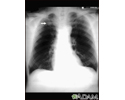
Coccidioidomycosis - chest X-ray - illustration
This chest x-ray shows the affects of a fungal infection, coccidioidomycosis. In the middle of the left lung (seen on the right side of the picture) there are multiple, thin-walled cavities (seen as light areas) with a diameter of 2 to 4 centimeters. To the side of these light areas are patchy light areas with irregular and poorly defined borders. Other diseases that may explain these x-ray findings include lung abscesses, chronic pulmonary tuberculosis, chronic pulmonary histoplasmosis, and others.
Coccidioidomycosis - chest X-ray
illustration
-
Pulmonary nodule - front view chest x-ray - illustration
This x-ray shows a single lesion (pulmonary nodule) in the upper right lung (seen as a light area on the left side of the picture). The nodule has distinct borders (well-defined) and is uniform in density. Tuberculosis (TB) and other diseases can cause this type of lesion.
Pulmonary nodule - front view chest x-ray
illustration
-
Disseminated coccidioidomycosis - illustration
Disseminated coccidioidomycosis is caused by breathing in the spores of a fungus found in desert regions. The infection spreads throughout the body and is especially common in immunosuppressed people. Antifungals may help but the death rate is very high.
Disseminated coccidioidomycosis
illustration
-
Fungus - illustration
Fungal infections are caused by microscopic organisms (fungi) that can live on the skin. They can live on the dead tissues of the hair, nails, and outer skin layers.
Fungus
illustration
-
Coccidioidomycosis - chest X-ray - illustration
This chest x-ray shows the affects of a fungal infection, coccidioidomycosis. In the middle of the left lung (seen on the right side of the picture) there are multiple, thin-walled cavities (seen as light areas) with a diameter of 2 to 4 centimeters. To the side of these light areas are patchy light areas with irregular and poorly defined borders. Other diseases that may explain these x-ray findings include lung abscesses, chronic pulmonary tuberculosis, chronic pulmonary histoplasmosis, and others.
Coccidioidomycosis - chest X-ray
illustration
-
Pulmonary nodule - front view chest x-ray - illustration
This x-ray shows a single lesion (pulmonary nodule) in the upper right lung (seen as a light area on the left side of the picture). The nodule has distinct borders (well-defined) and is uniform in density. Tuberculosis (TB) and other diseases can cause this type of lesion.
Pulmonary nodule - front view chest x-ray
illustration
-
Disseminated coccidioidomycosis - illustration
Disseminated coccidioidomycosis is caused by breathing in the spores of a fungus found in desert regions. The infection spreads throughout the body and is especially common in immunosuppressed people. Antifungals may help but the death rate is very high.
Disseminated coccidioidomycosis
illustration
-
Fungus - illustration
Fungal infections are caused by microscopic organisms (fungi) that can live on the skin. They can live on the dead tissues of the hair, nails, and outer skin layers.
Fungus
illustration
Review Date: 9/10/2022
Reviewed By: Jatin M. Vyas, MD, PhD, Associate Professor in Medicine, Harvard Medical School; Associate in Medicine, Division of Infectious Disease, Department of Medicine, Massachusetts General Hospital, Boston, MA. Also reviewed by David C. Dugdale, MD, Medical Director, Brenda Conaway, Editorial Director, and the A.D.A.M. Editorial team.


