
Patent foramen ovale
PFO; Congenital heart defect - PFOPatent foramen ovale (PFO) is a hole between the left and right atria (upper chambers) of the heart. This hole exists in everyone before birth, but most often closes shortly after being born. PFO is what the hole is called when it fails to close naturally after a baby is born.
Causes
A foramen ovale allows blood to go around the lungs. A baby's lungs are not used when it grows in the womb, so the hole does not cause problems in an unborn infant.
The opening is supposed to close soon after birth, but sometimes it does not. In about 1 out of 4 people, the opening never closes. If it does not close, it is called a PFO.
The cause of a PFO is unknown. There are no known risk factors. It can be found along with other heart abnormalities such as atrial septal aneurysms or Chiari network.
Symptoms
Infants with a PFO and no other heart defects do not have symptoms. Some adults with PFOs also suffer from migraine headaches.
Exams and Tests
An echocardiogram can be done to diagnose a PFO. If the PFO is not easily seen, a cardiologist can perform a "bubble test." Saline solution (salt water) is injected into the body as the cardiologist watches the heart on an ultrasound (echocardiogram) monitor. If a PFO exists, tiny air bubbles will be seen moving from the right to left side of the heart.
Echocardiogram
An echocardiogram is a test that uses sound waves to create pictures of the heart. The picture and information it produces is more detailed than a s...

Treatment
This condition is not treated unless there are other heart problems, symptoms, or if the person had a stroke caused by a blood clot to the brain.
Treatment most often requires a procedure called cardiac catheterization, which is performed by a trained cardiologist to permanently seal the PFO. Open heart surgery is no longer used to treat this condition unless another surgery is being performed.
Cardiac catheterization
Cardiac catheterization involves passing a thin flexible tube (catheter) into the right or left side of the heart. The catheter is most often insert...

Outlook (Prognosis)
An infant who has no other heart defects will have normal health and life span.
Possible Complications
Unless there are other defects, there are no complications from a PFO in most cases.
Some people may have a condition with shortness of breath and low arterial blood oxygen levels when sitting or standing. This is called platypnea-orthodeoxia. This is rare.
Rarely, people with PFOs may have a higher rate of a certain type of stroke (called paradoxical thromboembolic stroke). In a paradoxical stroke, a blood clot that develops in a vein (often leg veins) breaks free and travels to the right side of the heart. Normally, this clot would then continue to the lungs, but in someone with a PFO, the clot could pass through the hole to the left side of the heart. It may then be pumped out to the body, travel to the brain and become stuck there, preventing blood flow to that part of the brain (stroke).
Stroke
A stroke occurs when blood flow to a part of the brain stops. A stroke is sometimes called a "brain attack. " If blood flow is cut off for longer th...

Having a very mobile septum along with a PFO may lead to a higher risk for having a stroke.
Some people may take medicines to prevent blood clots.
When to Contact a Medical Professional
Contact your health care provider if your baby turns blue when crying or having a bowel movement, has difficulty feeding, or showing poor growth.
References
Kliegman RM, St Geme JW, Blum NJ, Shah SS, et al. Acyanotic congenital heart disease: left-to-right shunt lesions. In: Kliegman RM, St Geme JW, Blum NJ, Shah SS, Tasker RC, Wilson KM, eds. Nelson Textbook of Pediatrics. 21st ed. Philadelphia, PA: Elsevier; 2020:chap 453.
Therrien J, Marelli AJ. Congenital heart disease in adults. In: Goldman L, Cooney KA, eds. Goldman-Cecil Medicine. 27th ed. Philadelphia, PA: Elsevier; 2024:chap 55.
Valente AM, Dorfman AL, Babu-Narayan SV, Kreiger EV. Congenital heart disease in the adolescent and adult. In: Libby P, Bonow RO, Mann DL, Tomaselli GF, Bhatt DL, Solomon SD, eds. Braunwald's Heart Disease: A Textbook of Cardiovascular Medicine. 12th ed. Philadelphia, PA: Elsevier; 2022:chap 82.
-
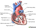
Heart - section through the middle - illustration
The interior of the heart is composed of valves, chambers, and associated vessels.
Heart - section through the middle
illustration
Review Date: 10/23/2023
Reviewed By: Michael A. Chen, MD, PhD, Associate Professor of Medicine, Division of Cardiology, Harborview Medical Center, University of Washington Medical School, Seattle, WA. Also reviewed by David C. Dugdale, MD, Medical Director, Brenda Conaway, Editorial Director, and the A.D.A.M. Editorial team.
 All rights reserved.