
Entropion
Eyelid - entropion; Eye pain - entropion; Tearing - entropionEntropion is the turning in of an edge of an eyelid. This causes the lashes to rub against the eye. It most often is seen on the lower eyelid.
Causes
Entropion can be present at birth (congenital).
In babies, it rarely causes problems because the lashes are very soft and do not easily damage the eye. In older people, the condition is most often caused by a spasm or weakening of the muscles surrounding the lower part of the eye.
Another cause can be trachoma infection, which can lead to scarring of the inner side of the lid. This is rare in North America and Europe. However, trachoma scarring is one of the three leading causes of blindness in the world.
Trachoma
Trachoma is an infection of the eye caused by bacteria called chlamydia.

Risk factors for entropion are:
- Aging
-
Chemical burn
Chemical burn
Chemicals that touch skin can lead to a reaction on the skin, throughout the body, or both.
 ImageRead Article Now Book Mark Article
 - Infection with trachoma
Symptoms
Symptoms include:
- Decreased vision if the cornea is damaged
- Excessive tearing
-
Eye discomfort or pain
Eye discomfort
Pain in the eye may be described as a burning, throbbing, aching, or stabbing sensation in or around the eye. It may also feel like you have a forei...
ImageRead Article Now Book Mark Article -
Eye irritation
Eye irritation
Eye burning with discharge is burning, itching, or drainage from the eye of any substance other than tears.
 ImageRead Article Now Book Mark Article
 - Redness
Exams and Tests
In most cases, your health care provider can diagnose this condition by looking at your eyelids. Special tests are not often necessary.
Treatment
Artificial tears can keep the eye from becoming dry and may help you feel better. Surgery to correct the position of the eyelids works well in most cases.
Outlook (Prognosis)
The outlook is most often good if the condition is treated before eye damage occurs.
Possible Complications
Dry eye and irritation may increase the risk for:
-
Corneal abrasions
Corneal abrasions
Corneal injury is a wound to the part of the eye known as the cornea. The cornea is the crystal clear (transparent) tissue that covers the front of ...
 ImageRead Article Now Book Mark Article
 -
Corneal ulcers
Corneal ulcers
The cornea is the clear tissue at the front of the eye. A corneal ulcer is an open sore in the outer layer of the cornea. It is often caused by inf...
ImageRead Article Now Book Mark Article - Eye infections
When to Contact a Medical Professional
Contact your provider if:
- Your eyelids turn inward.
- You constantly feel as though there is something in your eye.
Something in your eye
Eye emergencies include cuts, scratches, objects in the eye, burns, chemical exposure, and blunt injuries to the eye or eyelid. Certain eye infectio...
ImageRead Article Now Book Mark Article
If you have entropion, the following should be considered an emergency:
- Decreasing vision
-
Light sensitivity
Light sensitivity
Photophobia is eye discomfort in bright light.
ImageRead Article Now Book Mark Article - Pain
- Eye redness that increases rapidly
Prevention
Most cases cannot be prevented. Treatment reduces the risk of complications.
See your provider if you have red eyes after visiting an area where there is trachoma (such as North Africa or South Asia).
References
Cioffi GA, Liebmann JM. Diseases of the visual system. In: Goldman L, Cooney KA, eds. Goldman-Cecil Medicine. 27th ed. Philadelphia, PA: Elsevier; 2024:chap 391.
Gigantelli JW. Entropion. In: Yanoff M, Duker JS, eds. Ophthalmology. 6th ed. Philadelphia, PA: Elsevier; 2023:chap 12.5.
-
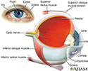
Eye - illustration
The eye is the organ of sight, a nearly spherical hollow globe filled with fluids (humors). The outer layer (sclera, or white of the eye, and cornea) is fibrous and protective. The middle layer (choroid, ciliary body and the iris) is vascular. The innermost layer (retina) is sensory nerve tissue that is light sensitive. The fluids in the eye are divided by the lens into the vitreous humor (behind the lens) and the aqueous humor (in front of the lens). The lens itself is flexible and suspended by ligaments which allow it to change shape to focus light on the retina, which is composed of sensory neurons.
Eye
illustration
-
Eye - illustration
The eye is the organ of sight, a nearly spherical hollow globe filled with fluids (humors). The outer layer (sclera, or white of the eye, and cornea) is fibrous and protective. The middle layer (choroid, ciliary body and the iris) is vascular. The innermost layer (retina) is sensory nerve tissue that is light sensitive. The fluids in the eye are divided by the lens into the vitreous humor (behind the lens) and the aqueous humor (in front of the lens). The lens itself is flexible and suspended by ligaments which allow it to change shape to focus light on the retina, which is composed of sensory neurons.
Eye
illustration
Review Date: 10/9/2024
Reviewed By: Linda J. Vorvick, MD, Clinical Professor, Department of Family Medicine, UW Medicine, School of Medicine, University of Washington, Seattle, WA. Also reviewed by David C. Dugdale, MD, Medical Director, Brenda Conaway, Editorial Director, and the A.D.A.M. Editorial team.
 All rights reserved.