Age-related macular degeneration
Macular degeneration is an eye disorder that slowly destroys sharp, central vision. This makes it difficult to see fine details and read.
The disease is most common in people over age 60, which is why it is often called age-related macular degeneration (ARMD or AMD).
Macular degeneration - Animation
The macula is the part of the retina that distinguishes fine details at the center of the field of vision. Macular degeneration results from a partial breakdown of the insulating layer between the retina and the choroid layer of blood vessels behind the retina. Macular degeneration results in the loss of central vision only.
Causes
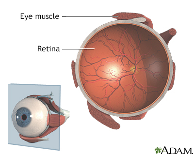
The retina is at the back of the eye. It changes light and images that enter the eye into nerve signals that are sent to the brain. A part of the retina called the macula makes vision sharper and more detailed. It is a yellow spot in the center of the retina. It has a high amount of two natural colors (pigments) called lutein and zeaxanthin.
AMD is caused by damage to the blood vessels that supply the macula. This change also harms the macula.
There are two types of AMD:
- Dry AMD occurs when the blood vessels under the macula become thin and brittle. Small yellow deposits, called drusen, form. Almost all people with macular degeneration start with the dry form.
- Wet AMD occurs in about 10% of people with macular degeneration. New abnormal and very fragile blood vessels grow under the macula. These vessels leak blood and fluid. This type of AMD causes most of the vision loss associated with the condition.
Health care providers are not sure what causes AMD. The condition is rare before age 55. It occurs mostly in people 75 years or older.
Risk factors for AMD are:
- Family history of AMD
- Being White
- Cigarette smoking
- High-fat diet
- Being female
Symptoms
You may not have any symptoms at first. As the disease gets worse, you may have problems with your central vision.
SYMPTOMS OF DRY AMD
The most common symptom of dry AMD is blurred vision. Objects in the center part of your vision often look distorted and dim, and colors look faded. You may have trouble reading print or seeing other details. But you can see well enough to walk and do most daily activities.
As dry AMD gets worse, you may need more light to read or do everyday tasks. A blurred spot in the center of vision gradually gets larger and darker.
In the later stages of dry AMD, you may not be able to recognize faces until they are close.
SYMPTOMS OF WET AMD
The most common early symptom of wet AMD is that straight lines look distorted and wavy.
There may be a small dark spot in the center of your vision that gets larger over time.
With both types of AMD, central vision loss can occur quickly. If this happens, you will need to be seen right away by an ophthalmologist. Make sure this eye doctor has experience in treating problems with the retina.
Exams and Tests
You will have an eye exam. Drops will be placed into your eyes to widen (dilate) your pupils. Your eye doctor will use special lenses to view your retina, blood vessels, and optic nerve.
Your eye doctor will look for specific changes in the macula and blood vessels and for drusen.
You may be asked to cover one eye and look at a pattern of lines called an Amsler grid. If the straight lines look wavy, it may be a sign of AMD. The American Macular Degeneration Foundation has an Amsler grid you can download along with instructions for how to check your vision.
Other tests that may be done include:
- Using special dye and camera to look at blood flow in the retina (fluorescein angiogram)
Fluorescein angiogram
Fluorescein angiography is an eye test that uses a special dye and camera to look at blood flow in the retina and choroid. These are the two layers ...
 ImageRead Article Now Book Mark Article
 - Taking a photo of the inner lining of the eye (fundus photography)
- Using light waves to view the retina (optical coherence tomography)
- A test that measures the pigment in the macula
Treatment
If you have advanced or severe dry AMD, no treatment can restore your vision.
If you have early AMD and do not smoke, a combination of certain vitamins, antioxidants, and zinc may prevent the disease from getting worse. But it cannot give you back vision that is already lost.
The combination is often called the "AREDS" formula. The supplements contain:
- 500 milligrams (mg) of vitamin C
- 400 international units (IU) of vitamin E
- 15 mg beta-carotene
- 80 mg of zinc
- 2 mg of copper
Only take this vitamin combination if your eye doctor recommends it. Make sure your eye doctor knows about any other vitamins or supplements you are taking. Smokers should not use this supplement.
AREDS may also benefit you if you have a family history and risk factors for AMD.
Lutein and zeaxanthin, which are substances found in green leafy vegetables, may also decrease your risk for age-related macular degeneration.
If you have wet AMD, your eye doctor may recommend:
- Laser surgery (laser photocoagulation) -- a small beam of light destroys the leaking, abnormal blood vessels.
Laser photocoagulation
Laser photocoagulation is eye surgery using a laser to shrink or destroy abnormal structures in the retina, or to intentionally cause scarring that c...
 ImageRead Article Now Book Mark Article
 - Photodynamic therapy -- a light activates a medicine that is injected into your body to destroy leaking blood vessels.
- Special medicines that prevent new blood vessels from forming in the eye are injected into the eye (this is an uncomfortable but painless process).
Low-vision aids (such as special lenses) and therapy can help you use the vision that you have more effectively, and improve your quality of life.
Low-vision aids
Low vision is a visual disability. Wearing regular glasses or contacts does not help. People with low vision have already tried the available medic...

Close follow-up with your eye doctor is important.
- For dry AMD, visit your eye doctor once a year for a complete eye exam.
- For wet AMD, you likely need frequent, perhaps monthly, follow-up visits.
Early detection of vision changes is important because the sooner you are treated, the better your outcome. Early detection leads to earlier treatment and often, a better outcome.
The best way to detect changes is by self-testing at home with the Amsler grid. Your eye doctor can give you a copy of the grid or you can print one from the Internet. Test each eye individually while wearing your reading glasses. If the lines look wavy, call your eye doctor right away for an appointment.
Treatments in development:
Stem cell research is showing some promise in restoring some vision in AMD. But this type of treatment is still years away at this point. Similarly, gene therapy may also have a place in the treatment of AMD. Levodopa, while most commonly used to treat Parkinson disease, may have a beneficial effect on wet AMD.
Support Groups
These resources may provide more information on macular degeneration:
- Macular Degeneration Association -- macularhope.org
- National Eye Institute -- www.nei.nih.gov/learn-about-eye-health/eye-conditions-and-diseases/age-related-macular-degeneration
Outlook (Prognosis)
AMD does not affect side (peripheral) vision. This means complete vision loss never occurs. AMD results in the loss of central vision only.
Mild, dry AMD usually does not cause disabling central vision loss.
Wet AMD often leads to significant vision loss.
In general, with AMD you may lose the ability to read, drive a car, and recognize faces at a distance. But most people with AMD can carry out daily tasks without much difficulty.
When to Contact a Medical Professional
If you have AMD, your eye doctor may recommend that you check your vision every day with an Amsler grid. Contact your provider right away if the lines look wavy. Also contact your eye doctor if you notice other changes in your vision.
Prevention
Although there is no known way to prevent macular degeneration, leading a healthy lifestyle can reduce your risk of developing AMD:
- Do not smoke
- Maintain a healthy diet that is high in fruits and vegetables and low in animal fat
- Exercise regularly
- Maintain a healthy weight
See your eye doctor regularly for dilated eye exams.
Reviewed By
Franklin W. Lusby, MD, Ophthalmologist, Lusby Vision Institute, La Jolla, CA. Also reviewed by David C. Dugdale, MD, Medical Director, Brenda Conaway, Editorial Director, and the A.D.A.M. Editorial team.
American Academy of Ophthalmology website. AAO PPP Retina/Vitreous Committee, Hoskins Center for Quality Eye Care. Preferred Practice Pattern Guideline. Age-related macular degeneration PPP 2019. www.aao.org/preferred-practice-pattern/age-related-macular-degeneration-ppp. Updated October 2019. Accessed August 7, 2023.
Chaikitmongkol V, Bressler NM, Bressler SB. Age-related macular degeneration: non-neovascular early AMD, intermediate AMD, and geographic atrophy. In: Sadda SR, Sarraf D, Freund B, et al, eds. Ryan's Retina. 7th ed. Philadelphia, PA: Elsevier; 2023:chap 66.
Jacoba CMP, Kim IK, Roh M. Age-related macular degeneration. In: Yanoff M, Duker JS, eds. Ophthalmology. 6th ed. Philadelphia, PA: Elsevier; 2023:chap 6.24.
Pugazhendhi A, Hubbell M, Jairam P, Ambati B. Neovascular macular degeneration: A review of etiology, risk factors, and recent advances in research and therapy. International Journal of Molecular Sciences. 2021;22(3):1170. PMID: 33504013 pubmed.ncbi.nlm.nih.gov/33504013/.


 All rights reserved.