
Meningococcal meningitis
Gram negative - meningococcusMeningitis is an infection of the membranes covering the brain and spinal cord. This covering is called the meninges.
Meningitis
Meningitis is an infection of the membranes covering the brain and spinal cord. This covering is called the meninges.

Bacteria are one type of germ that can cause meningitis. The meningococcal bacteria are one kind of bacteria that causes meningitis.
Causes
Meningococcal meningitis is caused by the bacteria Neisseria meningitidis (also known as meningococcus).
Meningococcus is the most common cause of bacterial meningitis in children and teens. It is a leading cause of bacterial meningitis in adults.
The infection occurs more often in winter or spring. It may cause local epidemics at boarding schools, college dormitories, or military bases.
Risk factors include recent exposure to someone with meningococcal meningitis, complement deficiency, use of eculizumab, spleen removal or a spleen that does not function, and exposure to cigarette smoking.
Symptoms
Symptoms usually come on quickly, and may include:
- Fever and chills
- Mental status changes
Mental status changes
Confusion is the inability to think as clearly or quickly as you normally do. You may feel disoriented and have difficulty paying attention, remembe...
 ImageRead Article Now Book Mark Article
 - Nausea and vomiting
- Purple, bruise-like areas (purpura)
Purpura
Purpura is purple-colored spots and patches that occur on the skin, and in mucus membranes, including the lining of the mouth.
 ImageRead Article Now Book Mark Article
 - Rash, pinpoint red spots (petechiae)
Rash
Rashes involve changes in the color, feeling or texture of your skin.
 ImageRead Article Now Book Mark Article
Petechiae
Bleeding into the skin can occur from broken blood vessels that form tiny red dots (called petechiae). Blood also can collect under the tissue in la...
 ImageRead Article Now Book Mark Article
 - Sensitivity to light (photophobia)
- Severe headache
- Stiff neck

Other symptoms that can occur with this disease:
- Agitation
- Bulging fontanelles in infants
Bulging fontanelles
A bulging fontanelle is an outward curving of an infant's soft spot (fontanelle).
 ImageRead Article Now Book Mark Article
 - Decreased consciousness
- Poor feeding or irritability in children
- Rapid breathing
Rapid breathing
Hyperventilation is rapid and deep breathing. It is also called overbreathing, and it may leave you feeling breathless.
 ImageRead Article Now Book Mark Article
 - Unusual posture with the head and neck arched backwards (opisthotonus)
Opisthotonus
Opisthotonos is a condition in which a person holds their body in an abnormal position. The person is usually rigid and arches their back, with thei...
 ImageRead Article Now Book Mark Article
Exams and Tests
Your health care provider will perform a physical exam. Questions will focus on symptoms and possible exposure to someone who might have the same symptoms, such as a stiff neck and fever.
If your provider thinks meningitis is possible, a lumbar puncture (spinal tap) will likely be done to obtain a sample of spinal fluid for testing.
Spinal tap
Cerebrospinal fluid (CSF) collection is a test to look at the fluid that surrounds the brain and spinal cord. CSF acts as a cushion, protecting the b...

Other tests that may be done include:
- Blood culture
Blood culture
A blood culture is a laboratory test to check for bacteria or other germs in a blood sample.
 ImageRead Article Now Book Mark Article
 - Chest x-ray
Chest x-ray
A chest x-ray is an x-ray of the chest, lungs, heart, large arteries, ribs, and diaphragm.
 ImageRead Article Now Book Mark Article
 - CT scan of the head
CT scan of the head
A head computed tomography (CT) scan uses many x-rays to create pictures of the head, including the skull, brain, eye sockets, and sinuses.
 ImageRead Article Now Book Mark Article
 - Complete blood count (CBC)
Complete blood count
A complete blood count (CBC) test measures the following:The number of white blood cells (WBC count)The number of red blood cells (RBC count)The numb...
 ImageRead Article Now Book Mark Article
 - Gram stain or, other special stains, and culture of the spinal fluid
Gram stain
A Gram stain is a test used to identify bacteria. It is one of the most common ways to quickly diagnose bacterial infection in the body.
ImageRead Article Now Book Mark ArticleCulture of the spinal fluid
A cerebrospinal fluid (CSF) culture is a lab test to look for bacteria, fungi, and viruses in the fluid that moves in the space around the spinal cor...
 ImageRead Article Now Book Mark Article
Treatment
Antibiotics will be started as soon as possible.
- Ceftriaxone is one of the most commonly used antibiotics.
- Penicillin in high doses can be effective for susceptible bacteria.
- If there is an allergy to penicillin, chloramphenicol may be used.
Sometimes, corticosteroids may be given.
People who have been in close contact with someone who has meningococcal meningitis should be given antibiotics to prevent infection.
Such people include:
- Household members
- Roommates in dormitories
- Military personnel who live in close quarters
- Those who come into close and long-term contact with an infected person
Outlook (Prognosis)
Early treatment improves the outcome. Death is possible. Young children and adults over age 50 have the highest risk of death.
Possible Complications
Long-term complications may include:
- Brain damage
- Hearing loss
- Buildup of fluid inside the skull that leads to brain swelling (hydrocephalus)
Hydrocephalus
Hydrocephalus is a buildup of fluid inside the skull that leads to the brain pushing against the skull. Hydrocephalus means "water on the brain. "...
ImageRead Article Now Book Mark Article - Buildup of fluid between the skull and brain (subdural effusion)
Subdural effusion
A subdural effusion is a collection of cerebrospinal fluid (CSF) trapped between the surface of the brain and the outer lining of the brain (the dura...
Read Article Now Book Mark Article - Inflammation of the heart muscle (myocarditis)
Myocarditis
Myocarditis is inflammation of the heart muscle. The condition is called pediatric myocarditis when it occurs in children.
 ImageRead Article Now Book Mark Article
 - Seizures
Seizures
A seizure is the physical changes in behavior that occurs during an episode of specific types of abnormal electrical activity in the brain. The term ...
 ImageRead Article Now Book Mark Article
When to Contact a Medical Professional
Call 911 or the local emergency number or go to an emergency room if you suspect meningitis in a young child who has the following symptoms:
- Feeding difficulties
- High-pitched cry
- Irritability
- Persistent unexplained fever
Meningitis can quickly become a life-threatening illness.
Prevention
Close contacts in the same household, school, or day care center should be watched for early signs of the disease as soon as the first person is diagnosed. All family and close contacts of this person should begin antibiotic treatment as soon as possible to prevent spread of the infection. Ask your provider about this during the first visit.
Always use good hygiene habits, such as washing hands before and after changing a diaper or after using the toilet.
Vaccines for meningococcus are effective for controlling spread and minimizing the severity of infection. They are currently recommended for:
Vaccines for meningococcus
All content below is taken in its entirety from the CDC Meningococcal Vaccine Information Statement (VIS): www. cdc. gov/vaccines/hcp/current-vis/men...

- Adolescents
- College students in their first year living in dormitories
- Military recruits
- Travelers to certain parts of the world
Although rare, people who have been vaccinated can still develop the infection.
References
Centers for Disease Control and Prevention website. Meningitis. About bacterial meningitis. www.cdc.gov/meningitis/about/bacterial-meningitis.html. Updated January 9, 2024. Accessed September 6, 2024.
Nath A. Meningitis: bacterial, viral, and other. In: Goldman L, Cooney KA, eds. Goldman-Cecil Medicine. 27th ed. Philadelphia, PA: Elsevier; 2024:chap 381.
Sadarangani M. Neisseria meningitidis (meningococcus). In: Kliegman RM, St. Geme JW, Blum NJ, et al, eds. Nelson Textbook of Pediatrics. 22nd ed. Philadelphia, PA: Elsevier; 2025:chap 237.
Stephens DS. Neisseria meningitidis. In: Bennett JE, Dolin R, Blaser MJ, eds. Mandell, Douglas, and Bennett's Principles and Practice of Infectious Diseases. 9th ed. Philadelphia, PA: Elsevier; 2020:chap 211.
The organs of the central nervous system (brain and spinal cord) are covered by connective tissue layers collectively called the meninges. Consisting of the pia mater (closest to the CNS structures), the arachnoid and the dura mater (farthest from the CNS), the meninges also support blood vessels and contain cerebrospinal fluid. These are the structures involved in meningitis, an inflammation of the meninges, which, if severe, may become encephalitis, an inflammation of the brain.
Meninges of the brain
illustration
The organs of the central nervous system (brain and spinal cord) are covered by 3 connective tissue layers collectively called the meninges. Consisting of the pia mater (closest to the CNS structures), the arachnoid and the dura mater (farthest from the CNS), the meninges also support blood vessels and contain cerebrospinal fluid. These are the structures involved in meningitis, an inflammation of the meninges, which, if severe, may become encephalitis, an inflammation of the brain.
Meninges of the spine
illustration
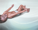
Meningococcemia is a life-threatening infection that occurs when the bacteria Neisseria meningitidis invades the blood stream. Bleeding into the skin (petechiae and purpura) usually occurs and the tissue may die (become necrotic or gangrenous). If the patient survives, the areas heal with scarring.
Meningococcal lesions on the back
illustration
The central nervous system comprises the brain and spinal cord. The peripheral nervous system includes nerves outside the brain and spinal cord.
Central nervous system and peripheral nervous system
illustration
CSF (cerebrospinal fluid) is a clear fluid that circulates in the space surrounding the spinal cord and brain. A CSF cell count is a test to measure the number of red and white blood cells that are in CSF.
CSF cell count
illustration
One of the physically demonstrable symptoms of meningitis is Brudzinski's sign. Severe neck stiffness causes a patient's hips and knees to flex when the neck is flexed.
Brudzinski's sign of meningitis
illustration
One of the physically demonstrable symptoms of meningitis is Kernig's sign. Severe stiffness of the hamstrings causes an inability to straighten the leg when the hip is flexed to 90 degrees.
Kernig's sign of meningitis
illustration


The organs of the central nervous system (brain and spinal cord) are covered by connective tissue layers collectively called the meninges. Consisting of the pia mater (closest to the CNS structures), the arachnoid and the dura mater (farthest from the CNS), the meninges also support blood vessels and contain cerebrospinal fluid. These are the structures involved in meningitis, an inflammation of the meninges, which, if severe, may become encephalitis, an inflammation of the brain.
Meninges of the brain
illustration
The organs of the central nervous system (brain and spinal cord) are covered by 3 connective tissue layers collectively called the meninges. Consisting of the pia mater (closest to the CNS structures), the arachnoid and the dura mater (farthest from the CNS), the meninges also support blood vessels and contain cerebrospinal fluid. These are the structures involved in meningitis, an inflammation of the meninges, which, if severe, may become encephalitis, an inflammation of the brain.
Meninges of the spine
illustration
Meningococcemia is a life-threatening infection that occurs when the bacteria Neisseria meningitidis invades the blood stream. Bleeding into the skin (petechiae and purpura) usually occurs and the tissue may die (become necrotic or gangrenous). If the patient survives, the areas heal with scarring.
Meningococcal lesions on the back
illustration
The central nervous system comprises the brain and spinal cord. The peripheral nervous system includes nerves outside the brain and spinal cord.
Central nervous system and peripheral nervous system
illustration
CSF (cerebrospinal fluid) is a clear fluid that circulates in the space surrounding the spinal cord and brain. A CSF cell count is a test to measure the number of red and white blood cells that are in CSF.
CSF cell count
illustration
One of the physically demonstrable symptoms of meningitis is Brudzinski's sign. Severe neck stiffness causes a patient's hips and knees to flex when the neck is flexed.
Brudzinski's sign of meningitis
illustration
One of the physically demonstrable symptoms of meningitis is Kernig's sign. Severe stiffness of the hamstrings causes an inability to straighten the leg when the hip is flexed to 90 degrees.
Kernig's sign of meningitis
illustration
Review Date: 8/29/2024
Reviewed By: Jatin M. Vyas, MD, PhD, Professor in Medicine, Harvard Medical School; Associate in Medicine, Division of Infectious Disease, Department of Medicine, Massachusetts General Hospital, Boston, MA. Also reviewed by David C. Dugdale, MD, Medical Director, Brenda Conaway, Editorial Director, and the A.D.A.M. Editorial team.
 All rights reserved.