Calcium pyrophosphate arthritis
Calcium pyrophosphate dihydrate (CPPD) arthritis is a joint disease that can cause attacks of arthritis. Like gout, crystals form in the joints. But in this arthritis, the crystals are not formed from uric acid.
Causes
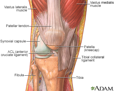
Deposition of calcium pyrophosphate dihydrate (CPPD) causes this form of arthritis. The buildup of this chemical forms crystals in the cartilage of joints. This leads to attacks of joint swelling and pain most often in the knees, wrists, ankles, and shoulders. Other joints can be affected as well. In contrast to gout, the metatarsal-phalangeal joint of the big toe is not affected.
Arthritis
Arthritis is inflammation or degeneration of one or more joints. A joint is the area where 2 bones meet. There are more than 100 different types of...

Joint swelling
Joint swelling is the buildup of fluid in the soft tissue surrounding the joint or the joint itself.

Among older adults, CPPD is a common cause of sudden (acute) arthritis in one joint. The attack may be caused by:
- Injury to the joint
- Medical stress
CPPD arthritis mainly affects the elderly because joint degeneration and osteoarthritis increases with age. Such joint damage increases the tendency of CPPD deposition. However, CPPD arthritis is also more common in people with:
Osteoarthritis
Osteoarthritis (OA) is the most common joint disorder. It is due to aging and wear and tear on a joint.

- Hemochromatosis
Hemochromatosis
Hemochromatosis is a condition in which there is too much iron in the body. It is also called iron overload.
 ImageRead Article Now Book Mark Article
 - Parathyroid gland disease
- Dialysis-dependent renal failure
Symptoms
In most cases, CPPD deposits do not cause any symptoms. Instead, x-rays of affected joints such as knees show characteristic deposits of calcium.
Some people with chronic CPPD deposits in large joints may have the following symptoms:
- Pain
- Swelling
- Warmth
- Redness
Attacks of joint pain can last for months. There may be no symptoms between attacks.
In some people CPPD arthritis causes severe damage to a joint.
CPPD arthritis can also occur in the upper and lower spine. Pressure on spinal nerves may cause pain in the arms or legs.
Because the symptoms are similar, CPPD arthritis can be confused with:
- Gouty arthritis (gout)
Gouty arthritis (gout)
Gout is a type of arthritis. It occurs when uric acid builds up in the blood and causes inflammation in the joints. Acute gout is a painful conditio...
 ImageRead Article Now Book Mark Article
 - Osteoarthritis
- Rheumatoid arthritis
Rheumatoid arthritis
Rheumatoid arthritis (RA) is a disease that leads to inflammation of the joints and surrounding tissues. It is a long-term disease. It can also aff...
 ImageRead Article Now Book Mark Article
Exams and Tests
Most arthritic conditions show similar symptoms. Carefully testing the joint fluid for crystals can help the health care provider diagnose the condition.
You may undergo the following tests:
- Joint fluid exam to detect white blood cells and calcium pyrophosphate crystals
- Joint x-rays to look for joint damage and calcium deposits in joint spaces
Joint x-rays
This test is an x-ray of a knee, shoulder, hip, wrist, ankle, or other joint.
 ImageRead Article Now Book Mark Article
 - Other joint imaging tests such as CT scan, MRI or ultrasound, if required
- Blood tests to screen for conditions that are linked to calcium pyrophosphate arthritis
Treatment
Treatment may involve removing fluid to relieve pressure in the joint. A needle is placed into the joint and fluid is aspirated. Some common treatment options are:
- Steroid injections: to treat severely swollen joints
- Oral steroids: to treat multiple swollen joints
- Nonsteroidal anti-inflammatory medicines (NSAIDs): to ease the pain
- Colchicine: to treat attacks of CPPD arthritis
- For chronic CPPD arthritis in multiple joints, methotrexate or hydroxychloroquine may be helpful
Outlook (Prognosis)
Most people do well with treatment to reduce the acute joint pain. A medicine such as colchicine may help prevent repeat attacks. There is no treatment to remove the CPPD crystals.
Possible Complications
Permanent joint damage can occur without treatment.
When to Contact a Medical Professional
Contact your provider if you have attacks of joint swelling and joint pain.
Prevention
There is no known way to prevent this disorder. However, treating other problems that may cause CPPD arthritis may make the condition less severe.
Regular follow-up visits may help prevent permanent damage of the affected joints.
Reviewed By
Neil J. Gonter, MD, Assistant Professor of Medicine, Columbia University, NY and private practice specializing in Rheumatology at Rheumatology Associates of North Jersey, Teaneck, NJ. Review provided by VeriMed Healthcare Network. Also reviewed by David C. Dugdale, MD, Medical Director, Brenda Conaway, Editorial Director, and the A.D.A.M. Editorial team.
Edwards NL. Crystal deposition diseases. In: Goldman L, Cooney KA, eds. Goldman-Cecil Medicine. 27th ed. Philadelphia, PA: Elsevier; 2024:chap 252.
Terkeltaub R. Calcium crystal disease: calcium pyrophosphate dihydrate and basic calcium phosphate. In: Firestein GS, Budd RC, Gabriel SE, Koretzky GA, McInnes IB, O'Dell JR, eds. Firestein & Kelley's Textbook of Rheumatology. 11th ed. Philadelphia, PA: Elsevier; 2021:chap 102.


 All rights reserved.