Colorectal cancer
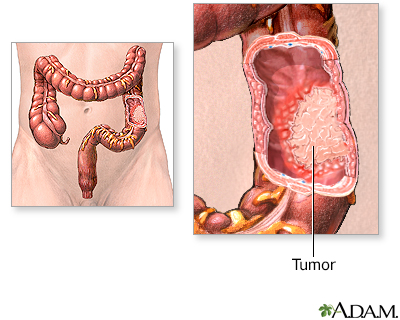
Colorectal cancer; Cancer - colon; Rectal cancer; Cancer - rectum; Adenocarcinoma - colon; Colon - adenocarcinoma; Colon carcinoma; Colon cancerColorectal cancer is cancer that starts in the large intestine (colon) or the rectum (end of the colon). It is also sometimes simply called colon cancer.
In the United States, colorectal cancer is one of the leading causes of deaths due to cancer. Early diagnosis can often lead to a complete cure.
Causes
Nearly all colorectal cancers begin as noncancerous (benign) lumps (polyps) in the lining of the colon and rectum. These can slowly develop into cancer.
Noncancerous (benign) lumps (polyps)
A colorectal polyp is a growth on the lining of the colon or rectum.

You have a higher risk for colorectal cancer if you:
- Are age 45 or older
- Drink alcohol
- Smoke tobacco
- Are overweight or have obesity
- Are African American or of eastern European descent
- Eat a lot of red or processed meats
- Eat a low-fiber and high-fat diet
- Have a diet low in fruits and vegetables
- Have colorectal polyps
Colorectal polyps
A colorectal polyp is a growth on the lining of the colon or rectum.
 ImageRead Article Now Book Mark Article
 - Have inflammatory bowel disease (Crohn disease or ulcerative colitis)
Crohn disease
Crohn disease is a disease where parts of the digestive tract become inflamed. It most often involves the lower end of the small intestine and the be...
 ImageRead Article Now Book Mark Article
Ulcerative colitis
Ulcerative colitis is a condition in which the lining of the large intestine (colon) and rectum become inflamed. It is a form of inflammatory bowel ...
 ImageRead Article Now Book Mark Article
 - Have a family history of colorectal cancer
Some inherited diseases also increase the risk of developing colorectal cancer. One of the most common is called Lynch syndrome.
Symptoms
Many cases of colon cancer have no symptoms. If there are symptoms, the following may indicate colon cancer:
-
Abdominal pain and tenderness in the lower abdomen
Abdominal pain and tenderness
Abdominal pain is pain that you feel anywhere between your chest and groin. This is often referred to as the stomach region or belly.
 ImageRead Article Now Book Mark Article
 -
Blood in the stool
Blood in the stool
Black or tarry stools with a foul smell are a sign of a problem in the upper digestive tract. It most often indicates that there is bleeding in the ...
 ImageRead Article Now Book Mark Article
 - Diarrhea, constipation, or other change in bowel habits
- Narrow stools
-
Weight loss with no known reason
Weight loss
Unexplained weight loss is a decrease in body weight, when you did not try to lose the weight on your own. Many people gain and lose weight. Uninten...
Read Article Now Book Mark Article
Exams and Tests
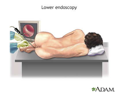
Through screening tests, colon cancer can be detected before symptoms develop. This is when the cancer is most curable. Abnormal stool screening tests should be followed up with a colonoscopy, which can see the entire colon.
Screening tests, colon cancer
Colon cancer screening can detect polyps and early cancers in the large intestine. This type of screening can find problems that can be treated befo...

Your health care provider will perform a physical exam and press on your belly area. The physical exam rarely shows any problems, although your provider may feel a lump (mass) in the abdomen. A rectal exam may reveal a mass in people with rectal cancer, but not colon cancer.
Colon culture
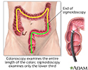
When polyps are discovered in a sigmoidoscopy (an inspection of the lower third of the large intestine), they are retrieved to be tested for cancer. If a large amount of polyps are found, a more thorough examination of the entire length of the large intestine (a colonoscopy) may be recommended.
Blood tests may be done for those diagnosed with colorectal cancer, including:
-
Complete blood count (CBC) to check for anemia
Complete blood count
A complete blood count (CBC) test measures the following:The number of white blood cells (WBC count)The number of red blood cells (RBC count)The numb...
 ImageRead Article Now Book Mark Article
 -
Liver function tests
Liver function tests
Liver function tests are common tests that are used to see how well the liver is working. Tests include:AlbuminAlpha-1 antitrypsinAlkaline phosphata...
 ImageRead Article Now Book Mark Article
If you are diagnosed with colorectal cancer, more tests will be done to see if the cancer has spread. This is called staging. CT or MRI scans of the abdomen, pelvic area, or chest may be used to stage the cancer. Sometimes, PET scans are also used.
Staging
Cancer staging is a way to describe how much cancer is in your body and where it is located. Staging helps determine where the original tumor is, ho...
Read Article Now Book Mark ArticleCT
A computed tomography (CT) scan is an imaging method that uses x-rays to create pictures of cross-sections of the body. Related tests include:Abdomin...

MRI
A magnetic resonance imaging (MRI) scan is an imaging test that uses powerful magnets and radio waves to create pictures of the body. It does not us...

PET
A positron emission tomography (PET) scan is a type of imaging test. It uses a radioactive substance called a tracer to look for disease in the body...
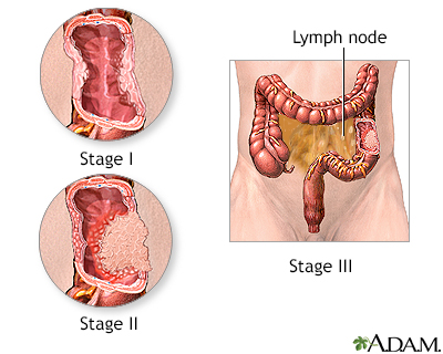
Read Article Now Book Mark ArticleStages of colorectal cancer are:
- Stage 0: Cancer is only on the innermost layer of the lining of the intestine
- Stage I: Cancer is in the inner layers of the colon
- Stage II: Cancer has spread through the muscle wall of the colon
- Stage III: Cancer has spread to the lymph nodes
- Stage IV: Cancer spread to other organs, such as the liver or lungs
Blood tests to detect tumor markers, such as carcinoembryonic antigen (CEA) may help your provider monitor your progress during and after treatment.
Carcinoembryonic antigen (CEA)
The carcinoembryonic antigen (CEA) test measures the level of CEA in the blood. CEA is a protein normally found in the tissue of a developing baby i...

Stages of cancer
The staging of a carcinoma has to do with the size of the tumor, and the degree to which it has penetrated. When the tumor is small and has not penetrated the mucosal layer, it is said to be stage I cancer. Stage II tumors are into the muscle wall, and stage III involves nearby lymph nodes. The rare stage IV cancer has spread (metastasized) to remote organs.
Treatment
Treatment depends on many things, including the stage of the cancer. Treatments may include:
- Endoscopic surgery (less invasive surgery using a lighted, flexible tube)
- Surgery
-
Chemotherapy
Chemotherapy
The term chemotherapy is used to describe cancer-killing drugs. Chemotherapy may be used to:Cure the cancerShrink the cancerPrevent the cancer from ...
 ImageRead Article Now Book Mark Article
 -
Radiation therapy
Radiation therapy
Radiation therapy uses high-powered radiation (such as x-rays or gamma rays), particles, or radioactive seeds to kill cancer cells.
 ImageRead Article Now Book Mark Article
 -
Immunotherapy
Immunotherapy
Immunotherapy is a type of cancer treatment that relies on the body's infection-fighting system (immune system). It uses substances made by the body...
Read Article Now Book Mark Article -
Targeted therapy
Targeted therapy
You are having a targeted therapy to try to kill cancer cells. You may receive targeted therapy alone or also have other treatments at the same time...
Read Article Now Book Mark Article
SURGERY
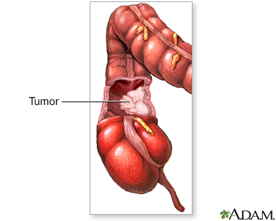
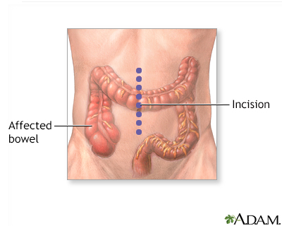
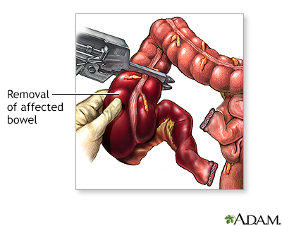
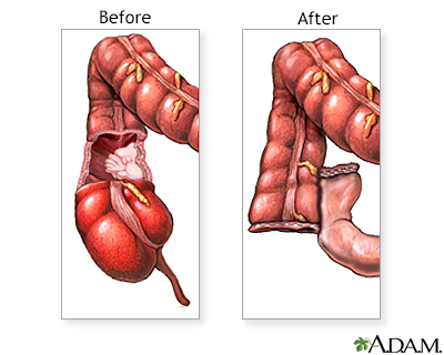
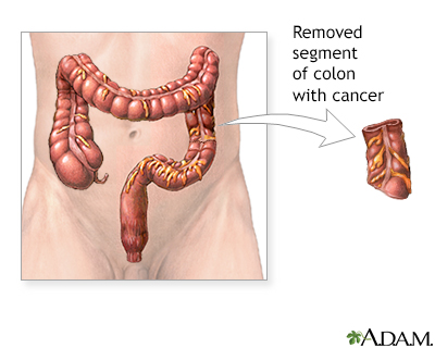
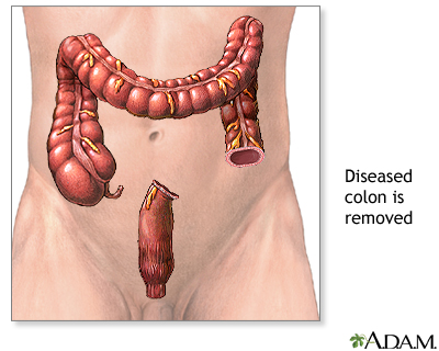
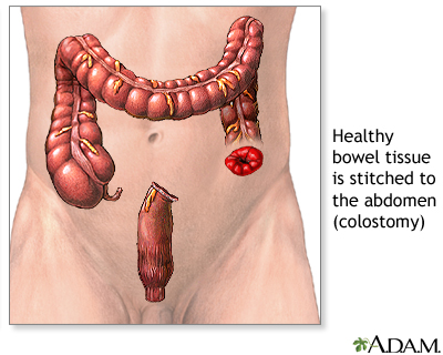
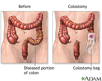
Stage 0 colon cancer may be treated by removing the tumor using endoscopic surgery (colonoscopy). For stages I, II, and III cancer, more extensive surgery is needed to remove all or part of the colon and rectum that is cancerous. This surgery is called colon resection (colectomy).
Colon resection
Large bowel resection is surgery to remove all or part of your large bowel. This surgery is also called colectomy. The large bowel is also called t...

CHEMOTHERAPY
Chemotherapy involves taking medicines that kill cancer cells. You may receive just one type of medicine or a combination of medicines.
Most people with stage III colon cancer receive chemotherapy after surgery for 3 to 6 months. This is called adjuvant chemotherapy. Even though the tumor was removed, chemotherapy is given to treat any cancer cells that may remain.
Chemotherapy is also used to improve symptoms and prolong survival in people with stage IV colon cancer.
IMMUNOTHERAPY
Immunotherapy involves taking medicines that increase the ability of your own immune system to destroy cancer cells. Immunotherapy has different side effects than chemotherapy.
RADIATION
Radiation therapy involves using radiation to kill cancer cells. Radiation therapy is often used in the treatment of rectal cancer.
TARGETED THERAPY
- Targeted treatment zeroes in on specific targets (molecules) in cancer cells. These targets play a role in how cancer cells grow and survive. Using these targets, the drug disables the cancer cells so they cannot spread. Targeted therapy may be given as pills or may be injected into a vein.
- You may have targeted therapy along with surgery, chemotherapy, or radiation treatment.
CANCER IN THE LIVER
For people with stage IV disease that has spread to the liver, treatment can be directed at the cancer tumors in the liver. This may include:
Spread to the liver
Liver metastases refer to cancer that has spread to the liver from somewhere else in the body. Liver metastases are not the same as cancer that start...

- Burning the liver tumors (ablation)
- Delivering chemotherapy or radiation directly into the liver
- Freezing the liver tumors (cryotherapy)
- Surgery
- Radioactive beads/spheres that deliver treatment to kill the cancer cells
- Alcohol (ethanol) injected into the liver tumors to kill cancer cells
Support Groups
You can ease the stress of the disease by joining a colon cancer support group. Sharing with others who have common experiences and problems can help you not feel alone.
Colon cancer support group
The following organizations are good resources for information on colorectal cancer:American Cancer Society -- www. cancer. org/cancer/colon-rectal-c...
Read Article Now Book Mark ArticleOutlook (Prognosis)
With treatment, stages 0, I, II, and III cancers often are cured, although higher stages of cancer are less likely to be cured. In most cases, stage IV cancer is not curable, but there are exceptions, including sometimes when the spread of the cancer is limited to the liver. In order for a person to be cured, treatment must get rid of all of the cancer. But there is a chance that the cancer will come back. If this occurs, curing the cancer is much less likely than before.
Cancer treatment can cause problems such as:
- Bowel obstruction from surgical scarring.
- Many sorts of short- and long-term side effects from chemotherapy, immunotherapy, radiation, and therapy targeted to the liver.
Possible Complications
Complications may include:
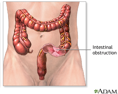
- Blockage of the colon, causing bowel obstruction
Bowel obstruction
Intestinal obstruction is a partial or complete blockage of the bowel. The contents of the intestine cannot pass through it.
 ImageRead Article Now Book Mark Article
 - Cancer returning in the colon
- Cancer spreading to other organs or tissues (metastasis)
Metastasis
Metastasis is the movement or spreading of cancer cells from one organ or tissue to another. Cancer cells usually spread through the blood or the ly...
 ImageRead Article Now Book Mark Article
 - Development of a second primary colorectal cancer
When to Contact a Medical Professional
Contact your provider if you have:
- Black, tar-like stools
- Blood during a bowel movement
- Change in bowel habits
- Unexplained weight loss
Prevention
Colon cancer can almost always be caught by colonoscopy in early stages, when it is most curable. All adults age 45 and older should have a colon cancer screening. How often you should have screening depends upon the test being used.
Colon cancer screening can often find polyps before they become cancerous. Removing these polyps may prevent colon cancer.
People with certain risk factors for colon cancer may need earlier testing (before age 45) or more frequent testing.
A healthy lifestyle also may help reduce your risk for colon cancer:
Healthy lifestyle
Like any illness or disease, cancer can occur without warning. Many factors that increase your cancer risk are beyond your control, such as your fam...

- Get regular physical activity.
- Don't smoke or use tobacco.
- Maintain a healthy weight.
- Eat a diet rich in fruits and vegetables and low in red and processed meats.
References
Centers for Disease Control and Prevention website. Reducing risk for colorectal cancer. www.cdc.gov/colorectal-cancer/prevention/. Updated June 20, 2024. Accessed August 27, 2024.
Garber JJ, Chung DC. Colonic polyps and polyposis syndromes. In: Feldman M, Friedman LS, Brandt LJ, eds. Sleisenger and Fordtran's Gastrointestinal and Liver Disease. 11th ed. Philadelphia, PA: Elsevier; 2021:chap 126.
Lawler M, Johnston B, Van Schaeybroeck S, et al. Colorectal cancer. In: Niederhuber JE, Armitage JO, Kastan MB, Doroshow JH, Tepper JE, eds. Abeloff's Clinical Oncology. 6th ed. Philadelphia, PA: Elsevier; 2020:chap 74.
National Cancer Institute website. Colorectal Cancer Prevention (PDQ) - Health Professional Version. www.cancer.gov/types/colorectal/hp/colorectal-prevention-pdq. Updated March 28, 2024. Accessed August 27, 2024.
National Comprehensive Cancer Network website. NCCN clinical practice guidelines in oncology (NCCN guidelines): colorectal cancer screening. Version 1.2024 - February 27, 2024. www.nccn.org/professionals/physician_gls/pdf/colorectal_screening.pdf. Updated February 27, 2024. Accessed August 27, 2024.
Patel SG, May FP, Anderson JC, et al. Updates on age to start and stop colorectal cancer screening: recommendations from the U.S. Multi-Society Task Force on Colorectal Cancer. Am J Gastroenterol. 2022;117(1):57-69. PMID: 34962727 pubmed.ncbi.nlm.nih.gov/34962727/.
Qaseem A, Crandall CJ, Mustafa RA, et al. Screening for colorectal cancer in asymptomatic average-risk adults: a guidance statement from the American College of Physicians. Ann Intern Med. 2019;171(9):643-654. PMID: 31683290 pubmed.ncbi.nlm.nih.gov/31683290/.
US Preventive Services Task Force website. Final recommendation statement. Colorectal cancer: screening. www.uspreventiveservicestaskforce.org/uspstf/recommendation/colorectal-cancer-screening. Published May 18, 2021. Accessed February 11, 2024.
-
Barium enema - illustration
The barium enema is a valuable diagnostic tool that helps detect abnormalities in the large intestine (colon). The barium enema, along with colonoscopy, remain standards in the diagnosis of colon cancer, ulcerative colitis, and other diseases of the colon.
Barium enema
illustration
-
Colonoscopy - illustration
There are 3 basic tests for colon cancer; a stool test (to check for blood), sigmoidoscopy (inspection of the lower colon), and colonoscopy (inspection of the entire colon). All 3 are effective in catching cancers in the early stages, when treatment is most beneficial.
Colonoscopy
illustration
-
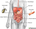
Digestive system - illustration
The esophagus, stomach, large and small intestine, aided by the liver, gallbladder and pancreas convert the nutritive components of food into energy and break down the non-nutritive components into waste to be excreted.
Digestive system
illustration
-
Rectal cancer - X-ray - illustration
A barium enema in a patient with cancer of the rectum.
Rectal cancer - X-ray
illustration
-
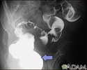
Sigmoid colon cancer - X-ray - illustration
A barium enema in a patient with cancer of the large bowel (sigmoid area).
Sigmoid colon cancer - X-ray
illustration
-
Spleen metastasis - CT scan - illustration
This CT scan of the upper abdomen shows multiple tumors in the liver and spleen that have spread (metastasized) from an original intestinal cancer (carcinoma).
Spleen metastasis - CT scan
illustration
-
Structure of the colon - illustration
The large intestine is a long hollow organ lined with mucous membrane (mucosa). Muscle layers wrap around the entire length and help move food material through to the rectum.
Structure of the colon
illustration
-
Stages of cancer - illustration
The staging of a carcinoma has to do with the size of the tumor, and the degree to which it has penetrated. When the tumor is small and has not penetrated the mucosal layer, it is said to be stage I cancer. Stage II tumors are into the muscle wall, and stage III involves nearby lymph nodes. The rare stage IV cancer has spread (metastasized) to remote organs.
Stages of cancer
illustration
-
Colon culture - illustration
When polyps are discovered in a sigmoidoscopy (an inspection of the lower third of the large intestine), they are retrieved to be tested for cancer. If a large amount of polyps are found, a more thorough examination of the entire length of the large intestine (a colonoscopy) may be recommended.
Colon culture
illustration
-
Colon cancer - Series
Presentation
-
Colostomy - Series
Presentation
-
Large bowel resection - Series
Presentation
-
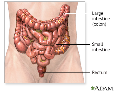
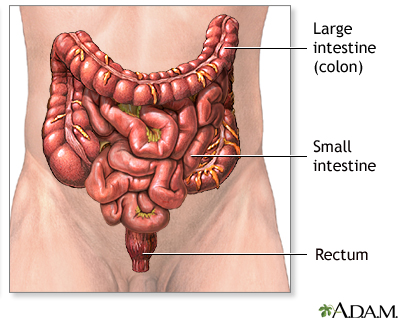
Large intestine (colon) - illustration
The large intestine is the portion of the digestive system most responsible for absorption of water from the indigestible residue of food. The ileocecal valve of the ileum (small intestine) passes material into the large intestine at the cecum. Material passes through the ascending, transverse, descending and sigmoid portions of the colon, and finally into the rectum. From the rectum, the waste is expelled from the body.
Large intestine (colon)
illustration
-
Barium enema - illustration
The barium enema is a valuable diagnostic tool that helps detect abnormalities in the large intestine (colon). The barium enema, along with colonoscopy, remain standards in the diagnosis of colon cancer, ulcerative colitis, and other diseases of the colon.
Barium enema
illustration
-
Colonoscopy - illustration
There are 3 basic tests for colon cancer; a stool test (to check for blood), sigmoidoscopy (inspection of the lower colon), and colonoscopy (inspection of the entire colon). All 3 are effective in catching cancers in the early stages, when treatment is most beneficial.
Colonoscopy
illustration
-
Digestive system - illustration
The esophagus, stomach, large and small intestine, aided by the liver, gallbladder and pancreas convert the nutritive components of food into energy and break down the non-nutritive components into waste to be excreted.
Digestive system
illustration
-
Rectal cancer - X-ray - illustration
A barium enema in a patient with cancer of the rectum.
Rectal cancer - X-ray
illustration
-
Sigmoid colon cancer - X-ray - illustration
A barium enema in a patient with cancer of the large bowel (sigmoid area).
Sigmoid colon cancer - X-ray
illustration
-
Spleen metastasis - CT scan - illustration
This CT scan of the upper abdomen shows multiple tumors in the liver and spleen that have spread (metastasized) from an original intestinal cancer (carcinoma).
Spleen metastasis - CT scan
illustration
-
Structure of the colon - illustration
The large intestine is a long hollow organ lined with mucous membrane (mucosa). Muscle layers wrap around the entire length and help move food material through to the rectum.
Structure of the colon
illustration
-
Stages of cancer - illustration
The staging of a carcinoma has to do with the size of the tumor, and the degree to which it has penetrated. When the tumor is small and has not penetrated the mucosal layer, it is said to be stage I cancer. Stage II tumors are into the muscle wall, and stage III involves nearby lymph nodes. The rare stage IV cancer has spread (metastasized) to remote organs.
Stages of cancer
illustration
-
Colon culture - illustration
When polyps are discovered in a sigmoidoscopy (an inspection of the lower third of the large intestine), they are retrieved to be tested for cancer. If a large amount of polyps are found, a more thorough examination of the entire length of the large intestine (a colonoscopy) may be recommended.
Colon culture
illustration
-
Colon cancer - Series
Presentation
-
Colostomy - Series
Presentation
-
Large bowel resection - Series
Presentation
-
Large intestine (colon) - illustration
The large intestine is the portion of the digestive system most responsible for absorption of water from the indigestible residue of food. The ileocecal valve of the ileum (small intestine) passes material into the large intestine at the cecum. Material passes through the ascending, transverse, descending and sigmoid portions of the colon, and finally into the rectum. From the rectum, the waste is expelled from the body.
Large intestine (colon)
illustration
Review Date: 4/18/2023
Reviewed By: John Roberts, MD, Professor of Internal Medicine (Medical Oncology), Yale Cancer Center, New Haven, CT. He is board certified in Internal Medicine, Medical Oncology, Pediatrics, Hospice and Palliative Medicine. Review provided by VeriMed Healthcare Network. Internal review and update on 02/20/2024 by David C. Dugdale, MD, Medical Director, Brenda Conaway, Editorial Director, and the A.D.A.M. Editorial team. Editorial update 08/27/2024.







![<strong>Large bowel resection - series</strong><p>The large bowel [large intestine or the colon] is part of the digestive system. It runs from the small intestine to the rectum. It is made up of three portions; the ascending, transverse and descending colon. The ascending colon is sometimes referred to as the right colon; the descending colon is sometimes referred to as the left, or sigmoid colon.</p> Large bowel resection - series](../../graphics/images/en/10255.jpg)
