CT angiography – chest
CT angiography combines a CT scan with the injection of dye. This technique is able to create pictures of the blood vessels in the chest and upper abdomen. CT stands for computed tomography.
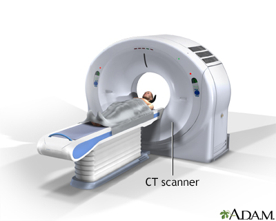
How the Test is Performed
You will be asked to lie on a narrow table that slides into the center of the CT scanner.
While inside the scanner, the machine's x-ray beam rotates around you.
A computer creates multiple separate images of the body area, called slices. These images can be stored, viewed on a monitor, or printed on film. Three-dimensional models of the chest area can be created by stacking the slices together.
You must be still during the exam, because movement causes blurred images. You may be told to hold your breath for short periods of time.
Complete scans usually take only a few minutes. The newest scanners can image your entire body, head to toe, in less than 30 seconds.
How to Prepare for the Test
Certain exams require a special dye, called contrast, to be delivered into the body before the test starts. Contrast helps certain areas show up better on x-rays.
- Contrast can be given through a vein (IV) in your hand or forearm. If contrast is used, you may also be asked not to eat or drink anything for 4 to 6 hours before the test.
- Let your health care provider know if you have ever had a reaction to contrast. You may need to take medications before the test in order to safely receive it.
- Before receiving the contrast, tell your provider if you take the diabetes medication metformin (Glucophage). You may need to take extra precautions.
The contrast can worsen kidney function problems in people with poorly functioning kidneys. Talk to your provider if you have a history of kidney problems.
Too much weight can damage the scanner. If you weigh more than 300 pounds (135 kilograms), talk to your provider about the weight limit before the test.
You will be asked to remove jewelry and wear a hospital gown during the study.
How the Test will Feel
The x-rays produced by the CT scan are painless. Some people may have discomfort from lying on the hard table.
If you have contrast through a vein, you may have a:
- Slight burning feeling
- Metallic taste in your mouth
- Warm flushing of your body
This is normal and usually goes away within a few seconds.
Why the Test is Performed
A chest CT angiogram may be done:
- For symptoms that suggest blood clots in the lungs, such as chest pain, rapid breathing, or shortness of breath
- After a chest injury or trauma
- Before surgery in the lung or chest
- To look for a possible site to insert a catheter for hemodialysis
- For swelling of the face or upper arms that cannot be explained
- To look for a suspected birth defect of the aorta or other blood vessels in the chest
- To look for a widening of an artery (aneurysm)
- To look for a tear in an artery (dissection)
- To assess certain blood vessels around the heart
Normal Results
Results are considered normal if no problems are seen.
What Abnormal Results Mean
A chest CT may show many disorders of the heart, lungs, or chest area, including:
- Suspected blockage of the superior vena cava: This large vein moves blood from the upper half of the body to the heart.
Blockage of the superior vena cava
SVC obstruction is a narrowing or blockage of the superior vena cava (SVC), which is the second largest vein in the human body. The superior vena ca...
 ImageRead Article Now Book Mark Article
 - Blood clot(s) in the lungs.
Blood clot(s) in the lungs
A pulmonary embolus is a blockage of an artery in the lungs. The most common cause of the blockage is a blood clot.
 ImageRead Article Now Book Mark Article
 - Abnormalities of the blood vessels in the lungs or chest, such as aortic arch syndrome.
Aortic arch syndrome
The aortic arch is the top part of the main artery carrying blood away from the heart. Aortic arch syndrome refers to a group of signs and symptoms ...
ImageRead Article Now Book Mark Article - Aortic aneurysm (in the chest area), also called thoracic aortic aneurysm.
Aortic aneurysm (in the chest area)
Aortic dissection is a serious condition in which there is a tear in the wall of the major artery carrying blood out of the heart (aorta). As the te...
 ImageRead Article Now Book Mark Article
 - Narrowing of part of the major artery leading out of the heart (aorta).
Major artery leading out of the heart (...
The aorta is a larger artery that carries blood from the heart to the vessels that supply the rest of the body with blood. If part of the aorta is n...
 ImageRead Article Now Book Mark Article
 - Tear in the wall of an artery (dissection).
- Inflammation of the blood vessel walls (vasculitis).
- Problems with pulmonary vessels, including narrowing of pulmonary veins of abnormal connections between pulmonary arteries and veins (arteriovenous malformations).
Risks
Risks of CT scans include:
- Being exposed to radiation
- Allergic reaction to contrast dye
- Damage to kidneys from contrast dye
CT scans use more radiation than regular x-rays. Having many x-rays or CT scans over time may increase your risk for cancer. However, the risk from any one scan is small. You and your provider should weigh this risk against the benefits of getting a correct diagnosis for a medical problem. Most modern scanners use techniques to use less radiation.
Some people have allergies to contrast dye. Let your provider know if you have ever had an allergic reaction to injected contrast dye.
- The most common type of contrast given into a vein contains iodine. If you have an iodine allergy, you may have nausea or vomiting, sneezing, itching, or hives if you get this type of contrast.
Nausea or vomiting
Nausea is feeling an urge to vomit. It is often called "being sick to your stomach. "Vomiting or throwing-up forces the contents of the stomach up t...
 ImageRead Article Now Book Mark Article
Sneezing
A sneeze is a sudden, forceful, uncontrolled burst of air through the nose and mouth.
 ImageRead Article Now Book Mark Article
Itching
Itching is a tingling or irritation of the skin that makes you want to scratch the area. Itching may occur all over the body or only in one location...
 ImageRead Article Now Book Mark Article
Hives
Hives are raised, often itchy, red bumps (welts) on the surface of the skin. They can be an allergic reaction to food or medicine. They can also ap...
 ImageRead Article Now Book Mark Article
 - If you absolutely must be given such contrast, your provider may give you antihistamines (such as Benadryl) and/or steroids before the test.
- The kidneys help remove iodine out of the body. Those with kidney disease or diabetes may need to receive extra fluids after the test to help flush the iodine out of the body.
Rarely, the dye may cause a life-threatening allergic response called anaphylaxis. If you have any trouble breathing during the test, you should notify the scanner operator immediately. Scanners come with an intercom and speakers, so someone can hear you at all times.
Anaphylaxis
Anaphylaxis is a life-threatening type of allergic reaction.

Your provider may ask you to avoid the use of metformin for two days after the CT-angiogram.
Reviewed By
Frank D. Brodkey, MD, FCCM, Associate Professor, Section of Pulmonary and Critical Care Medicine, University of Wisconsin School of Medicine and Public Health, Madison, WI. Also reviewed by David C. Dugdale, MD, Medical Director, Brenda Conaway, Editorial Director, and the A.D.A.M. Editorial team.
Carmichael SP, Mowery NT, Martin RS, Meredith JW. Management of acute trauma. In: Townsend CM Jr, Beauchamp RD, Evers BM, Mattox KL, eds. Sabiston Textbook of Surgery. 21st ed. Philadelphia, PA: Elsevier; 2022:chap 17.
Gilman M. Congenital and developmental diseases of the lungs and airways. In: Digumarthy SR, Abbara S, Chung JH, eds. Problem Solving in Chest Imaging. Philadelphia, PA: Elsevier; 2020:chap 15.
Reekers JA. Angiography: principles, techniques and complications. In: Adam A, Dixon AK, Gillard JH, Schaefer-Prokop CM, eds. Grainger & Allison's Diagnostic Radiology: A Textbook of Medical Imaging. 7th ed. Philadelphia, PA: Elsevier; 2021:chap 78.
 All rights reserved.