
Leg CT scan
CAT scan - leg; Computed axial tomography scan - leg; Computed tomography scan - leg; CT scan - legA computed tomography (CT) scan of the leg makes cross-sectional pictures of the leg. It uses x-rays to create the images.
How the Test is Performed
You will lie on a narrow table that slides into the center of the CT scanner.
Once you are inside the scanner, the machine's x-ray beam rotates around you. Modern "spiral" scanners can perform the exam without stopping.
A computer creates separate images of the body area, called slices. These images can be stored, viewed on a monitor, or printed on film. Three-dimensional (3D) models of the leg can be created by adding the slices together.
You will need to lie still during the exam. Movement can cause blurred images. You may need to hold your breath for short periods of time.
The scan should take only 10 to 15 minutes.
How to Prepare for the Test
Some exams use a special dye, called contrast that is put into your body before the test starts. Contrast helps certain areas show up better on the x-rays.
- Contrast can be given through a vein (IV) in your hand or forearm. If contrast is used, you may also be asked not to eat or drink anything for 4 to 6 hours before the test.
- Let your health care provider know if you have ever had a reaction to contrast. You may need to take medicines before the test in order to avoid this problem.
- Before having the contrast, tell your provider if you take the diabetes medicine metformin (Glucophage). You may need to take extra steps before the test if you take this drug.
Too much weight can cause damage to the scanner's working parts. Find out if the CT machine has a weight limit if you weigh more than 300 pounds (135 kilograms).
You will wear a hospital gown during the study. You will need to take off all jewelry as metal can affect the CT images.
How the Test will Feel
Some people may be uncomfortable lying on the hard table.
Contrast given through an IV may cause a slight burning feeling, a metal taste in the mouth, and a warm flushing of the body. These feelings are normal and go away in a few seconds.
Why the Test is Performed
CT scan makes detailed pictures of the body very quickly. The test may help look for:
- An abscess or infection
Abscess
An abscess is a collection of pus in any part of the body. In most cases, the area around an abscess is swollen and inflamed.
 ImageRead Article Now Book Mark Article
 - A mass that is felt during a physical exam
- The cause of pain or other problems in the foot, ankle, or knee joints (usually when an MRI cannot be done)
MRI
A magnetic resonance imaging (MRI) scan is an imaging test that uses powerful magnets and radio waves to create pictures of the body. It does not us...
 ImageRead Article Now Book Mark Article
 - A broken bone
- Masses and tumors, including cancer
- Healing problems or scar tissue following surgery
- A deformity that can be evaluated to help planning for surgery
A CT scan may also be used to guide a surgeon to the right area during a biopsy.
Biopsy
A biopsy is the removal of a small piece of tissue for lab examination.

Normal Results
Results are considered normal if the leg being examined looks OK.
What Abnormal Results Mean
Abnormal results may be due to:
- Degenerative changes due to age
- Abscess or infection
- Blood clot in the leg (deep venous thrombosis)
Blood clot in the leg
Deep vein thrombosis (DVT) is a condition that occurs when a blood clot forms in a vein deep inside a part of the body. DVT mainly affects the large...
 ImageRead Article Now Book Mark Article
 - Broken or fractured bone
Broken
If more pressure is put on a bone than it can stand, it will split or break. A break of any size is called a fracture. If the broken bone punctures...
 ImageRead Article Now Book Mark Article
 - Cancer
Cancer
Cancer is the uncontrolled growth of abnormal cells in the body. Cancerous cells are also called malignant cells.
Read Article Now Book Mark Article - Damage to the knee, foot, or ankle joint
- Noncancerous bone tumor
- Healing problems or development of scar tissue after surgery
Risks
Risks of CT scans include:
- Being exposed to radiation
- Allergic reaction to contrast dye
- Birth defect if done during pregnancy
CT scans expose you to more radiation than regular x-rays. Having many x-rays or CT scans over time may raise your risk for cancer, but the risk from any one scan is small. Talk to your provider about this risk against the benefits of the test.
x-rays
X-rays are a type of electromagnetic radiation, just like visible light. An x-ray machine sends individual x-ray waves through the body. The images...
Some people have allergies to contrast dye. Let your provider know if you have ever had this type of reaction.
- The most common type of contrast given into a vein contains iodine. A person with an iodine allergy may have nausea or vomiting, sneezing, itching, or hives from this type of contrast.
- If you must have this type of contrast, you may get antihistamines (such as Benadryl) or steroids before the test.
- The kidneys help remove iodine out of the body. People with kidney disease or diabetes may need to get extra fluids after the test to help flush the iodine out of the body.
Diabetes
Diabetes is a long-term (chronic) disease in which the body cannot regulate the amount of sugar in the blood.
 ImageRead Article Now Book Mark Article
The dye may cause a life-threatening allergic response called anaphylaxis. This is rare. Tell the scanner operator right away if you have any trouble breathing during the test. Scanners come with an intercom and speakers, so the operator can hear you at all times.
Anaphylaxis
Anaphylaxis is a life-threatening type of allergic reaction.

References
Kapoor G, Toms AP. Current status of imaging of the musculoskeletal system. In: Adam A, Dixon AK, Gillard JH, Schaefer-Prokop CM, eds. Grainger & Allison's Diagnostic Radiology. 7th ed. Philadelphia, PA: Elsevier; 2021:chap 38.
Yepuri N, Pruekprasert N, Cooney RN. Surgical complications. In: Townsend CM Jr, Beauchamp RD, Evers BM, Mattox KL, eds. Sabiston Textbook of Surgery. 21st ed. St Louis, MO: Elsevier; 2022:chap 12.
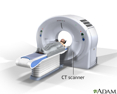
CT scan - illustration
CT stands for computerized tomography. In this procedure, a thin X-ray beam is rotated around the area of the body to be visualized. Using very complicated mathematical processes called algorithms, the computer is able to generate a 3-D image of a section through the body. CT scans are very detailed and provide excellent information for the physician.
CT scan
illustration
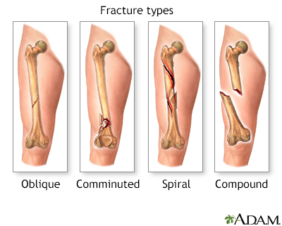
Fracture types (1) - illustration
There are several types of bone fracture, including oblique -- a fracture which goes at an angle to the axis, comminuted -- a fracture of many relatively small fragments, spiral -- a fracture which runs around the axis of the bone, and compound -- a fracture (also called open) which breaks the skin.
Fracture types (1)
illustration
Leg skeletal anatomy - illustration
The lower leg is comprised of two bones, the tibia and the smaller fibula. The thigh bone, or femur, is the large upper leg bone that connects the lower leg bones (knee joint) to the pelvic bone (hip joint).
Leg skeletal anatomy
illustration


CT scan - illustration
CT stands for computerized tomography. In this procedure, a thin X-ray beam is rotated around the area of the body to be visualized. Using very complicated mathematical processes called algorithms, the computer is able to generate a 3-D image of a section through the body. CT scans are very detailed and provide excellent information for the physician.
CT scan
illustration
Fracture types (1) - illustration
There are several types of bone fracture, including oblique -- a fracture which goes at an angle to the axis, comminuted -- a fracture of many relatively small fragments, spiral -- a fracture which runs around the axis of the bone, and compound -- a fracture (also called open) which breaks the skin.
Fracture types (1)
illustration
Leg skeletal anatomy - illustration
The lower leg is comprised of two bones, the tibia and the smaller fibula. The thigh bone, or femur, is the large upper leg bone that connects the lower leg bones (knee joint) to the pelvic bone (hip joint).
Leg skeletal anatomy
illustration
Review Date: 4/24/2023
Reviewed By: C. Benjamin Ma, MD, Professor, Chief, Sports Medicine and Shoulder Service, UCSF Department of Orthopaedic Surgery, San Francisco, CA. Also reviewed by David C. Dugdale, MD, Medical Director, Brenda Conaway, Editorial Director, and the A.D.A.M. Editorial team.
 All rights reserved.