Browse A-Z

More <
bookmarks-menu

Cone biopsy

Show Alternative Names
Biopsy - cone
Cervical conization
CKC
Cervical intraepithelial neoplasia - cone biopsy
CIN - cone biopsy
Precancerous changes of the cervix - cone biopsy
Cervical cancer - cone biopsy
Squamous intraepithelial lesion - cone biopsy
LSIL - cone biopsy
HSIL - cone biopsy
Low-grade cone biopsy
High-grade cone biopsy
Carcinoma in situ-cone biopsy
CIS - cone biopsy
ASCUS - cone biopsy
Atypical glandular cells - cone biopsy
AGUS - cone biopsy
Atypical squamous cells - cone biopsy
Pap smear - cone biopsy
HPV - cone biopsy
Human papilloma virus - cone biopsy
Cervix - cone biopsy
Colposcopy - cone biopsy

A cone biopsy (conization) is surgery to remove a sample of abnormal tissue from the cervix. The cervix is the lower part of the uterus (womb) that opens at the top of the vagina. Abnormal changes in the cells on the surface of the cervix is called cervical dysplasia.

How the Test is Performed

This procedure is done in the hospital or in a surgery center. During the procedure:

  • You will be given general anesthesia (asleep and pain-free), or medicines to help you relax and feel sleepy.
  • You will lie on a table and place your feet in stirrups to position your pelvis for exam. The health care provider will place an instrument (speculum) into your vagina to better see the cervix.
  • A small cone-shaped sample of tissue is removed from the cervix. The procedure may be performed using a wire loop heated by electrical current (LEEP procedure), a scalpel (cold knife biopsy), or a laser beam.
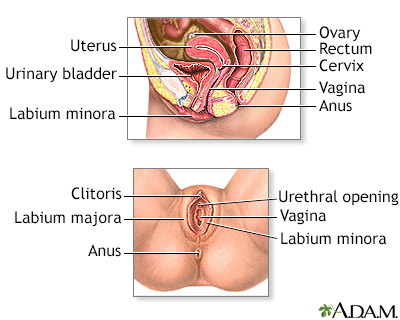
  • The cervical canal above the cone biopsy may also be scraped to remove cells for evaluation. This is call an endocervical curettage (ECC).
  • The sample is examined under a microscope for signs of cancer. This biopsy may also be a treatment if the provider removes all of the diseased tissue.

Most of the time, you will be able to go home the same day as the procedure.

How to Prepare for the Test

You may be asked to not eat or drink for 6 to 8 hours before the test.

How the Test will Feel

After the procedure, you may have some cramping or discomfort for about a week. For about 4 to 6 weeks avoid:

  • Douching (douching should never be done)
  • Sexual intercourse
  • Using tampons

For 2 to 3 weeks after the procedure, you may have discharge that is:

  • Bloody
  • Heavy
  • Yellow-colored

Why the Test is Performed

Cone biopsy is done to detect cervical cancer or early changes that lead to cancer. A cone biopsy is done if a test called colposcopy cannot find the cause of an abnormal Pap smear.

Cone biopsy may also be used to treat:

  • Moderate to severe types of abnormal cell changes (called CIN II or CIN III)
  • Very early stage cervical cancer (stage 0 or IA1)

Cervical cancer - Animation

Worldwide, cervical cancer is the third most common type of cancer in women. Luckily, it's much less common in the United States due to women receiving recommended routine Pap smears, the test designed to find cervical cancer sometimes even before abnormal cells turn to cancer. Cervical cancer starts in the cells on the surface of the cervix, the lower portion of the uterus. There are two types of cells on the surface of the cervix, squamous and columnar. Most cervical cancers come from these squamous cells. The cancer usually starts very slowly as a condition called dysplasia. This precancerous condition can be detected by Pap smear and is 100% treatable. Undetected, precancerous changes can develop into cervical cancer and spread to the bladder, intestines, lungs, and liver. It can take years for these precancerous changes to turn into cervical cancer. However, patients with cervical cancer do not usually have problems until the cancer is advanced and has spread. Most of the time, early cervical cancer has no symptoms. Symptoms of advanced cancer may include back pain, bone fractures, fatigue, heavy vaginal bleeding, urine leakage, leg pain, loss of appetite, and pelvic pain. If after having a Pap smear, the doctor finds abnormal changes on the cervix, a colposcopy can be ordered. Using a light and a low-powered microscope, the doctor will view the cervix under magnification. The doctor may remove pieces of tissue, called a biopsy, and send the sample to a laboratory for testing. If the woman is diagnosed with cervical cancer, the doctor will order more tests to determine how far the cancer has spread. This is called Staging. Treatment will depend on the stage of the cancer, the size and shape of the tumor, the woman's age and general health, and her desire to have children in the future. Early cervical cancer can be treated with surgery just to remove abnormal tissue, freeze abnormal cells, or burn abnormal tissue. Treatment for more advanced cervical cancer may include radical hysterectomy, removal of the uterus and much of the surrounding tissue, including lymph nodes and the upper part of the vagina. Radiation may be used to treat cancer that has spread beyond the pelvis, or if cancer returns. The woman may also have chemotherapy to kill cancer cells. Almost all cervical cancers are caused by human papilloma virus, or HPV. This common virus is spread through sexual intercourse. HPV vaccines can prevent infection. Practicing safe sex also reduces the risk of getting HPV. But, keep in mind most women diagnosed with cervical cancer have not had their regular Pap smears. Because Pap smears can find precancerous growths that are 100% treatable, it's very important for women to get Pap smears at regular intervals.

Normal Results

A normal result means there are no precancerous or cancerous cells in the cervix.

What Abnormal Results Mean

Most often, abnormal results mean that there are precancerous or cancerous cells in the cervix. These changes are called cervical intraepithelial neoplasia (CIN). The changes are divided into 3 groups:

  • CIN I -- mild dysplasia
  • CIN II -- moderate to marked dysplasia
  • CIN III -- severe dysplasia to carcinoma in situ

Abnormal results may also be due to cervical cancer.

Risks

Risks of cone biopsy include:

  • Bleeding
  • Incompetent cervix (which may lead to premature delivery)
  • Infection
  • Scarring of the cervix (which may cause painful periods, premature delivery, and difficulty getting pregnant)
  • Damage to the bladder or rectum

Cone biopsy may also make it hard for your provider to interpret abnormal Pap smear results in the future.

Review Date: 1/10/2022

Reviewed By

John D. Jacobson, MD, Department of Obstetrics and Gynecology, Loma Linda University School of Medicine, Loma Linda, CA. Also reviewed by David Zieve, MD, MHA, Medical Director, Brenda Conaway, Editorial Director, and the A.D.A.M. Editorial team.

References

Cohen PA, Jhingran A, Oaknin A, Denny L. Cervical cancer. Lancet. 2019;393(10167):169-182. PMID: 30638582 pubmed.ncbi.nlm.nih.gov/30638582/.

Salcedo MP, Phoolcharoen N, Schmeler KM. Intraepithelial neoplasia of the lower genital tract (cervix, vagina, vulva): etiology, screening, diagnosis, management. In: Gershenson DM, Lentz GM, Valea FA, Lobo RA, eds. Comprehensive Gynecology. 8th ed. Philadelphia, PA: Elsevier; 2022:chap 29.

Watson LA. Cervical conization. In: Fowler GC, ed. Pfenninger and Fowler's Procedures for Primary Care. 4th ed. Philadelphia, PA: Elsevier; 2020:chap 128.

Disclaimer

The information provided herein should not be used during any medical emergency or for the diagnosis or treatment of any medical condition. A licensed medical professional should be consulted for diagnosis and treatment of any and all medical conditions. Links to other sites are provided for information only -- they do not constitute endorsements of those other sites. No warranty of any kind, either expressed or implied, is made as to the accuracy, reliability, timeliness, or correctness of any translations made by a third-party service of the information provided herein into any other language. © 1997- A.D.A.M., a business unit of Ebix, Inc. Any duplication or distribution of the information contained herein is strictly prohibited.

All
Videos
Images
Cervical cancer

Cervical cancer

Animation

Female reproductive anatomy - Illustration Thumbnail

Female reproductive anatomy

Internal structures of the female reproductive anatomy include the uterus, ovaries, and cervix. External structures include the labium minora and majora, the vagina and the clitoris.

Illustration

Cold cone biopsy - Illustration Thumbnail

Cold cone biopsy

The cold cone biopsy is a surgical procedure requiring general anesthesia and is indicated by the presence of precancerous changes in the cervix.

Illustration

Cold cone removal - Illustration Thumbnail

Cold cone removal

In the cold cone removal procedure, a small cone-shaped sample of tissue is removed from the cervix and examined under a microscope for any signs of cancer.

Illustration

 
Cervical cancer

Cervical cancer

Animation

 
Female reproductive anatomy - Illustration Thumbnail

Female reproductive anatomy

Internal structures of the female reproductive anatomy include the uterus, ovaries, and cervix. External structures include the labium minora and majora, the vagina and the clitoris.

Illustration

Cold cone biopsy - Illustration Thumbnail

Cold cone biopsy

The cold cone biopsy is a surgical procedure requiring general anesthesia and is indicated by the presence of precancerous changes in the cervix.

Illustration

Cold cone removal - Illustration Thumbnail

Cold cone removal

In the cold cone removal procedure, a small cone-shaped sample of tissue is removed from the cervix and examined under a microscope for any signs of cancer.

Illustration

 
##RemoveMe##
 

Cervical cancer - Animation

Worldwide, cervical cancer is the third most common type of cancer in women. Luckily, it's much less common in the United States due to women receiving recommended routine Pap smears, the test designed to find cervical cancer sometimes even before abnormal cells turn to cancer.

Cervical cancer starts in the cells on the surface of the cervix, the lower portion of the uterus. There are two types of cells on the surface of the cervix, squamous and columnar. Most cervical cancers come from these squamous cells.

The cancer usually starts very slowly as a condition called dysplasia. This precancerous condition can be detected by Pap smear and is 100% treatable. Undetected, precancerous changes can develop into cervical cancer and spread to the bladder, intestines, lungs, and liver. It can take years for these precancerous changes to turn into cervical cancer. However, patients with cervical cancer do not usually have problems until the cancer is advanced and has spread.

Most of the time, early cervical cancer has no symptoms. Symptoms of advanced cancer may include back pain, bone fractures, fatigue, heavy vaginal bleeding, urine leakage, leg pain, loss of appetite, and pelvic pain.

If after having a Pap smear, the doctor finds abnormal changes on the cervix, a colposcopy can be ordered. Using a light and a low-powered microscope, the doctor will view the cervix under magnification. The doctor may remove pieces of tissue, called a biopsy, and send the sample to a laboratory for testing.

If the woman is diagnosed with cervical cancer, the doctor will order more tests to determine how far the cancer has spread. This is called Staging. Treatment will depend on the stage of the cancer, the size and shape of the tumor, the woman's age and general health, and her desire to have children in the future.

Early cervical cancer can be treated with surgery just to remove abnormal tissue, freeze abnormal cells, or burn abnormal tissue. Treatment for more advanced cervical cancer may include radical hysterectomy, removal of the uterus and much of the surrounding tissue, including lymph nodes and the upper part of the vagina. Radiation may be used to treat cancer that has spread beyond the pelvis, or if cancer returns. The woman may also have chemotherapy to kill cancer cells.

Almost all cervical cancers are caused by human papilloma virus, or HPV. This common virus is spread through sexual intercourse. HPV vaccines can prevent infection. Practicing safe sex also reduces the risk of getting HPV.

But, keep in mind most women diagnosed with cervical cancer have not had their regular Pap smears. Because Pap smears can find precancerous growths that are 100% treatable, it's very important for women to get Pap smears at regular intervals.


© 1997-ADAM Company Logo All rights reserved.